Download
1 / 35
400 likes | 870 Views
Mechanisms of heart failure. Definition of heart failure. Heart failure is a complex clinical syndrome that can result from any cardiac disorder that impairs the ability of the ventricle to eject blood. The cardinal manifestations of heart failure are dyspnoea and fatigue

E N D
Mechanisms of heart failure • Definition of heart failure Heart failure is a complex clinical syndrome that can result from any cardiac disorder that impairs the ability of the ventricle to eject blood. The cardinal manifestations of heart failure are dyspnoea and fatigue (which may limit exercise tolerance) and fluid retention (Which may lead to pulmonary and peripheral oedema). Both abnormalities impair the functional capacity and quality of life of affected individuals. Consensus recommendations for the management of chronic heart failure ACTION HF - AJC Jan 21,1999 vol 83(2A) - Packer M et al
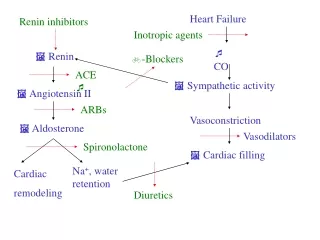
Definition of heart failure "Heart failure occurs when an abnormality of cardiac function causes the heart to fail to pump blood at a rate required by the metabolizing tissues or when the heart can do so only with an elevated filling pressure. The heart's inability to pump a sufficient amount of blood to meet the needs of the body tissues may be due to insufficient or defective cardiac filling and/or impaired contraction and emptying. Compensatory mechanisms increase blood volume and raise cardiac filling pressures, heart rate, and cardiac muscle mass to maintain the heart's pumping function and cause redistribution of blood flow”. National Heart, Lung and Blood Institute
Mechanisms of heart failure • Reduced volume of blood delivered to the systemic arterial bed. • One or both ventricles has elevated filling pressures. • Result: Retention of sodium and water in the intravascular and intersititial compartments. • Dyspnoea and oedema
Symptoms of heart failure • Dyspnoea - breathlessness - Increased awareness of respiration or difficulty in breathing . • If due to cardiac causes it is usually due to left ventricular failure and pulmonary congestion. • Pulm. capillary Hypertension • Restrictive ventilatory defect -VC and TV reduced • Lungs are stiffer -increased work of breathing • Air trapping - earlier closure of dependent airways • Airways resistance increased - congestion of peripheral airways. • V/Q mismatch - hypoxaemia • Increased ventilatory drive • Stretch receptors pulm. vessels & interstitium • hypoxia & acidosis • Incr. Work of breathing & impaired perfusion of resp. musc (low CO) - fatigue - dyspnoea
Symptoms of heart failure • Orthopnoea: Dyspnoea present when recumbent and relieved by elevation. No. of pillows. • Mech: Reduced pooling of fluid in lower extremities and abd. Increased venous return: Failing L Ventricle (flat portion of depressed F-S curve) - cannot accept extra volume - Increased pulmonary venous pressure - Pulmonary oedema
Symptoms of heart failure • Paroxysmal nocturnal dyspnoea: • Patient awakes suddenly with feeling of anxiety and suffocation - sits upright and gasps for breath. • Bronchspasm (Wheezing - “cardiac asthma”) • Congestion bronchial mucosa • Compression of small bronchi by interstitial pulmonary. oedema. • Increased work of breathing.
Symptoms of heart failure • Pulmonary oedema: • Increased pulmonary venous pressure - (failing LV) • Increased pulmonary capillary pressures. • Interstitial pulmonary oedema • Reduced pulmonary compliance • Increased airway resistance • Dyspnoea.
Pulmonary oedema: Braunwald: Heart disease –p463
Symptoms of heart failure Reduced Exercise Capacity • Dyspnoea - Pulmonary vascular congestion • Insufficient blood flow to exercising muscles • Inadequate augmentation of CO with exercise. • Impaired vasodilatation • Abnormal skeletal muscle. Metabolism • Deconditioning skeletal& respiratory muscles • Anxiety • Grade ‘cardiac status’ : NYHA 1-4 - Degree of exertion - Determine if a change has occurred.
Symptoms of heart failure • Fatique and weakness -Poor perfusion of skeletal muscles. • Impaired vasodilatation • Abnormal skeletal musc. Metabolism • sodium depletion / hypovolaemia / beta blockers. • Urinary symptoms • Nocturia - redistribution blood flow to kidneys at night. • Cerebral symptoms • Confusion, memory impairment, insomnia, disorientation, etc
Symptoms of heart failure Congestive Symptoms“Forward vs. Backward failure” • Fluid localizes behind the chamber initially affected. • Pressure in the venous and capillary bed behind the failing ventricle rises - Transudation of fluid into the interstitial bed • Fluid retention • Reduced GFR • Activation of RAAS
Symptoms of heart failure Congestive Symptoms“Forward vs. Backward failure • Left ventricle: Pulmonary congestion / oedema • Right ventricle: • Raised Jugular Venous Pressure • Hepatic congestion • Splancnic ooedema and ascites • Pleural effusion • Ankle oedema
Pathophysiological mechanisms that causes raised filling pressures and/ poor tissue perfusion: HF • Reduced cardiac contraction • An increased cardiac load • Valvular dysfunction • Diastolic dysfunction • High output states
Pathophysiological mechanisms that causes raised filling pressures and/ poor tissue perfusion: HF • Primary abnormality of the heart muscle - Cardiomyopathies/myocarditis • Coronary atherosclerosis - Ishaemia and infarction of the muscle • Longstanding excessive haemodynamic burden i.e valvular abnormality • causing myocardial damage Reduced cardiac contraction: “myocardial failure” Systolic dysfunction with a depressed LV ejection fraction (usually <40%) Generally accompanied by an increase in left ventricular end-diastolic and end systolic volumes
Pathophysiological mechanisms that causes raised filling pressures and/ poor tissue perfusion: HF • Adaptive mechanisms • The Frank Starling mechanism -Increased preload helps to • sustain cadiac performance • Myocardial hyperthophy • Neuro-hormonal actvation - to maintain arterial pressure and • perfusion vital organs. Vasoconstriction and • fluid and water retention Reduced cardiac contraction: “myocardial failure”
Pathophysiological mechanisms that causes raised filling pressures and/ poor tissue perfusion: HF The Frank Starling mechanism -Increased preload helps to sustain cadiac performance
Pathophysiological mechanisms that causes raised filling pressures and/ poor tissue perfusion: HF • An increased cardiac load • Cardiac output = Stroke vol x heart rate • Pre-load Contractility Afterload • Preload: Tension of the myocardial fibers at the end of diastole (degree of stretch): Venous filling pressure. • Afterload: Myocardial wall tension developed during systolic ejection: LV: resistance of aortic valve, peripheral vascular resistance and elasticity of major blood vessels. Laplace: T= PR/2xwall thickness. Ventricular wall tension is increased by ventricular dilatation, incr. intra-ventricular pressure or reduction in wall thickness
Pathophysiological mechanisms that causes raised filling pressures and/ poor tissue perfusion: HF • An increased cardiac load • Preload: • HF: Ejection fraction reduced – increase in volume blood remaining after systole – increase in diastolic volume and venous pressure. • Depression of the ventricular function curve: • Slight myocardial depression: CO maintained by increase in venous pressure (diastolic volume) Starling’s law and HR. • More severe myocardial dysfunction – large incr. in venous pressure – systemic and pulm. oedema. CO at rest may still be normal but fails to incr. with exercise • Severe HF: Decr. CO at rest: CO redistributed to vital organs
Pathophysiological mechanisms that causes raised filling pressures and/ poor tissue perfusion: HF • Afterload: • Systemic and pulmonary resistance • Physical characteristics of the vessel walls • The volume of blood that is ejected. Increase in after load decreases CO with an increase in end-diastolic volume which in turn increases afterload (Laplace) • Examples (LV) • Aortic stenosis • Hypertension • Elderly (Compliance vessels) • Conditions which causesventricular dilatation, incr. Intra-ventricular pressure or reduction in wall thickness: see Laplace – conditions that cause volume overload – I.e Aortic and mitral regurgitation, dilated cardiomyopathies etc
Mechanisms of load induced effects on cardiac performance • Myocardial remodelling in heart failure: • Geometric remodelling • Change in myocardial gene expression • Contractile proteiens (Myosin heavy chains), Na-K-ATPase, • Ca-ATPase,Beta 1 adrenoreceptors • Abnormal calcium homeostasis • Prolongation of the calcium current in association with • prolongation of contraction and relaxation • (Decr. Sarcolemmm Ca-ATPase activty etc.) • Apoptosis. • Programmed cell death – initiated cytokines, free radicals etc.
Mechanisms of load induced effects on cardiac performance • Myocardial remodelling in heart failure: • Geometric remodelling: • Ventricular hypertrophy - compensatory mechanism of increased load: • Increase in size of cells, mitochondria, myofibrils,interstitial collagen • Stimulus for hypertrophy: • Pressure overload: • Systolic wall stress increases • Parallel replication of myofibrils: • Thickening of myocytes: • Concentric hypertrophy • Volume overload: • Diastolic wall stress increases • Sarcomeres replicates in series • Elongation of myocytes • Ventricular dilatation / eccentric hypertrophy
Pathophysiological mechanisms that causes raised filling pressures and/ poor tissue perfusion: HF The High Output States • Low out-put failure (commonest) • Impaired peripheral circulation with systemic vasoconstriction and shunting of blood to the vital organs • Cold, pale / cyanotic extremities. • Pulse pressure may narrow • High output failure: Heart is required to pump abnormally large quantities of blood to deliver the required quota of oxygen to metabolizing tissues • Reduced vascular resistance, increased vascular capacitance and blood volume • Extremities are warm and flushed, pulse pressure may be wide • Arterial-mixed venous oxygen difference normal or reduced due to delivery of large amounts of arterial blood to non-metabolizing tissues.
Systolic Failure:The heart does not deliver the quantity of oxygen required by the metabolizing tissues. High output Low output Cardiac output: Rest N -high Low - N Exercise Fail to rise normally Fail to rise normally Arterial-mixed venous oxygen difference Low (? N rest) High (? N rest) (Admixture of blood diverted from metabolizing tissue) Peripheral circulation Blood volume Increased Vascular resistance Reduced Increased Vascular capacitance Increased Extremities Warm/flushed Cold/pale/cyanotic Pulse pressure Widens Narrows High output versus low output states
Pathophysiological mechanisms that causes raised filling pressures and/ poor tissue perfusion: HF • Causes: • Hyperthyroidism • Thyroid hormone: Direct effect on cardiac contractility and • Metabolism. Increased metabolic demands and decr. SVR • Aneamia (O2 delivery = blood flow x Hb x A –V sat): • Tissue hypoxia, decr. Blood viscosity, decr. SVR, incr. CO • Beriberi • Thiamine deficiency impairs pyruvate dehydrog. • Accumulation lactate and pyruvate, periheral vasodilatation, • decr. SVR, Incr CO. (also impairs myocardial metabolism) • Pregnancy • Arteriovenous fistulas Decreased SVR • Paget’s disease The high output states
Pathophysiological mechanisms that causes raised filling pressures and/ poor tissue perfusion: HF Valvular Dysfunction • Mitral stenosis: LA pressure incr., pulm. venous congestion, pulm. arterial hypertension, R heart failure. • Mitral regurgitation: LA dilates and pressure rises (compliance), LV dilates (vol. overload - proportion of CO regurgitated) – CO increases. LV dysfunction: Pulm. venous congestion due to mitral regurgitation and LV failure. • Aortic stenosis: Obstruction to LV outflow, LV hypertrophy (concentric – pressure overload), relative LV ischaemia, LV dysfunction: LV end-diastolic pressures and LA pressures rise, pulm. congestion. • Aortic regurgitation: Proportion of LV EF regurgitated, LV Dilates (volume overload) and CO increases. Diastolic pressure declines and coronary flow decreases. Dilated LV – incr. myocardial O2 demand. LV dysfunction -LV end-diastolic pressures and LA pressures rise, pulm. congestion.
Pathophysiological mechanisms that causes raised filling pressures and/ poor tissue perfusion: HF Diastolic dysfunction Altered ventricular relaxation:(isovolumetric relax. & early vent filling phases) • Dynamic process • Uptake Ca sarcoplasmic reticulum & Ca efflux from myocyte • Sarcoplasmic retic Ca ATPase and sarcolemmal Ca pumps. • Energy consuming
Diastolic dysfunction Altered Ventricular Filling • Early ventricular filling: Myocardium lenghtens rapidly and in-homogeneously • diastolic asynergy: Regional variation in onset rate and extent of lengthening • diastolic asynchrony: temporal dispersion. • End-diastolic filling • Myocardial elasticity: Change muscle length for change in force • Ventricular compliance: Change in volume for change in pressure • Ventricular stiffness: Inverse of compliance
Diastolic dysfunction Increased chamber stiffness • Rise in filling pressure. (steeper portion of pressure volume curve) • Volume overload: Acute valvular regurgitation / Acute LV failure - myocarditis. • Steeper ventricular pressure volume curve: Increase ventricular mass / wall thickness (hypertrophy) or intrinsic stiffness (infiltration, fibrosis, ischaemia) • Pressure volume curve displaced parallel upwards (Decreased ventricular distensibility: Extrinsic compression of ventricle. - constrictive pericarditis.
Diastolic dysfunction Effects of ventricular interaction Ventricles anatomically interlinked Systolic ventricular interaction Septum part of load against which each ventricle must work LV hypertrophy includes septum: R ventricle must work harder and becomes hypertrophied Diastolic ventricular interaction: Bernheim effect/reverse Volume overloading of one ventricle impairs the filling/function of the other ventricle
Left Heart Failure Leads to Right heart failure • Left Ventricular Failure Causes Elevation of: • Left Ventricular diastolic • Left Atrial • Pulmonary venous pressures • Backwards transmission of pressure • Protective mechanism against Pulmonary Oedema: • Increased lymphatic drainage • Capillary/alveolar barrier thickened and less permeable • Constriction of pulm. resistance vessels • Pulmonary vasoconstriction / Increased pulmonary vasc. resistance • Pulmonary hypertension • Ultimately Right Ventricular failure.
Right Heart Failure due to Pulmonary Disease (Cor Pulmonale) • Chronic Bronchitis and Emphysema: • Hypoxia induced pulmonary vasoconstriction. • Vasoconstrictive effect of hydrogen ions • Pulmonary artery pressure correlates • inversely with O2 sat • directly with PCO2 • Muscular hypertrophy of pulmonary arterioles • Increased blood viscosity - Increased hematocrit. • Interstitial Pulm. Fibrosis / Vasculitides: • Reduction in cross sectional area of pulm. vasc. bed