Download
1 / 36
360 likes | 596 Views
ABDOMEN. PREMED H&P. INSPECTION. 1. Patient should lie down on examination table (Supine Position) 2. Drape a sheet over the pubic region 3. Abdomen should be exposed from above the xiphoid process (just below the breasts) to the symphysis pubis

E N D
ABDOMEN PREMED H&P
1. Patient should lie down on examination table (Supine Position) • 2. Drape a sheet over the pubic region • 3. Abdomen should be exposed from above the xiphoid process (just below the breasts) to the symphysis pubis • 4. Patient arms should be at sides or folded across the chest
Appearance of Abdomen: • A) Flat or Distended or Protrubent? • B) Symmetry • C) Surgical scars / Striae / Dilated Veins /Other skin abnormalities • D) Protrusions
Umbilicus • Peristalsis – e.g. Intestinal Obstruction causes increased peristalsis • Pulsations - Aortic pulsation is visible in the epigastrium e.g. Aortic Aneurysm (Increased pulse) • Patient’s Movement
Performed before Percussion or Palpation • Above maneuvers can alter the frequency of bowel sounds (Palpating the abdomen may disturb the intestines) • Auscultate for normal bowel sounds – Place the stethoscope on the abdomen and listen for 15 to 20 seconds • 1) Presence of bowel sounds • 2) Listen carefully and assess for Frequency & Character
Absent bowel sounds – e.g. Ileus – Intestinal obstruction, eg. Peritonitis • Hyperactive bowel sounds – e.g. Inflammation of mucosa due to infections causing diarrhea • Bruits – E.g. Renal artery stenosis due to hypertension • Bruits can be heart at aorta, iliac and femoral arteries as well
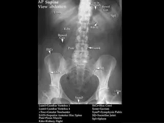
Aorta Renal Artery Iliac Artery Femoral Artery
Technique that has to be mastered with practice • Percuss in all four quadrants to assess for the following sounds: • A) Tympany • B) Dullness • Tympany (drum-like sounds) occur with gas in the abdomen • Dullness occurs with solid structures or fluid in the abdomen
Tympanic – Protrubent abdomen or intestinal obstruction • Dullness – e.g. Ascites, Feces, mass or *enlarged organ • During this procedure, make note of any pain that may indication inflammation or peritonitis
Two major organs that are percussable: • A) Liver • B) Spleen • Area covering liver Dull under percussion • **Size of liver must be determined by percussion • Splenomegaly Enlargement will be dull under percussion (Assesed by Palpation)
Liver is located deep to the ribs 7- 11 • Start at the right upper quadrant • Place the left hand behind the patient • Place your right hand on the mid-clavicular line below the lower border of liver felt as dullness • Press gently in and up while you push posteriorly • Asking the patient to take deep breaths while pushing up and down can be easier to palpate the liver edges
Hepatomegaly or Liver enlargement – e.g. Infections such as Malaria, Chronic abuse of alcohol
In a small percentage of adults, spleen is palpable • A palpable spleen can be felt by • Using the left hand to reach over posteriorly around the patient for support • Placing your right hand below the left costal margin.
What is the costovertebral angle? • Acute angle between the 12th rib and the vertebral column • To palpate the left kidney, place the right hand below the patient towards the costovertebral angle • Place the left hand gently in the left upper quandrant asking the patient to take deep breaths • Palpate firmly and deeply below the left costal margin • Kidney enlargement – e.g. Hydrophrosis, tumors, cysts
Assess Kindey Tenderness – “Costovertebral angle tenderness” • To assess – Place ball of one hand on the costovertebral angle and strike with the ulnar surface of the other hand making a “fist”: • Kidney tenderness – e.g. Pyelonephritis or kidney infection