abdomen
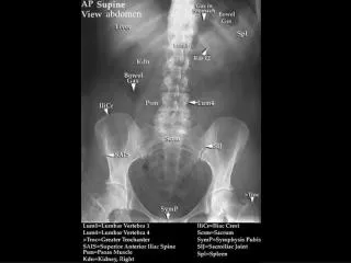
abdomen. AP Abdomen. Naso-gastric (NG tube). Iodonated contrast in Left kidney. Inferior vena cava filter. AP View UGI. Oblique View UGI. Small Bowel Follow Through (Early). Ligament Of Treitz. Duodenal c-loop. Jejunum. Pancreatic head. Small Bowel Follow Through (1 hour later).

abdomen
E N D
Presentation Transcript
AP Abdomen Naso-gastric (NG tube) Iodonated contrast in Left kidney Inferior vena cava filter
Small Bowel Follow Through (Early) Ligament Of Treitz Duodenal c-loop Jejunum Pancreatic head
Small Bowel Follow Through (1 hour later) jejunum Ilieum
AP Abdomen Naso-gastric (NG tube) Iodonated contrast in Left kidney Inferior vena cava filter
Operative Cholangiogram Gallbladder with gallstones Common bile duct Duodenal c loop Laparoscope
5 Nuclear Medicine-Biliary Tract Scan 2 • Gallbladder • Common bile duct • Duodenal loop • Right lobe of liver • Left lobe of liver 1 4 3
Intravenous Pyleogram Opacified renal parenchyma Minor calyx Renal pelvis Renal pelvis Renal papilla Inferior Inferior major calyx Peristaltic contraction Bladder
Intravenous Pyleogram Sacroilliac joint Sacroilliac joint Typical shape of female pelvis Contrast in distal ureter Body of uterus bladder phleblolith
Aortagram Splenic Artery Hepatic Artery Right renal artery Left renal artery
Aortagram Aorta Aorta Left renal arteries Right renal arteries Lumbar arteries Lumbar arteries Common illiac arteries Common illiac arteries
Aortagram Intercostal artery 1 2 Superior mesenteric artery
Superior Mesenteric Arteriogram A C • Middle colic artery • Superior mesenteric artery • Replaced right hepatic artery • Spinous process of L1 • Right Colic artery • Superior mesenteric artery • Ileocolic artery • Ileal (instestinal) arteries • Jejunal (instestinal) arteries B D E I I G H F H
Magnetic Resonance Angiogram Heart Renal Artery Left Kidney Aorta Right Kidney
Lumbararteries LeftRenal Arteriogram Left renal artery
Inferior Vena Cavagram Inferior Vena Cava Right external iliac vein Right common femoral vein
IS IS IS= Ischial Spine
Lymphangiogram Opacifying external illiac lymph nodes
AP Pelvic Arteriogram • Abdominal Aorta • Internal illiac artery • External illiac artery • Lumbar artery • Common femoral artery • Common illiac 4 6 5
RA RA RA= Rectus Abdominus