Working with ECGs
Working with ECGs. Dr Cynthia Lim Dr Dean Pritchard FACEMs, Emergency Department The Northern Hospital. ECG 123s. Measurement of electrical flow across the heart using electrodes placed on the chest and limbs

Working with ECGs
E N D
Presentation Transcript
Working with ECGs Dr Cynthia Lim Dr Dean Pritchard FACEMs, Emergency Department The Northern Hospital
ECG 123s • Measurement of electrical flow across the heart using electrodes placed on the chest and limbs • Deviation of electrical flow from normal pathways indicates cardiac anomaly or cardiac disease
The Leads • Limb Leads • aVR – Right arm • aVL – Left arm • aVF – Left leg • Vectors • Flow of +ve current • I – R arm L arm • II – R arm L leg • III – L arm L leg
The Leads • Chest leads • V1 • V2 • V3 • V4 • V5 • V6
Axis Look at leads I and avF If in left quadrant then look at lead II
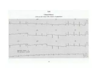
Pick the Problem… NORMAL ECG
ECG of 2 year old – normal or abnormal? Higher rate, Partial RBBB pattern, Dominant R V1, R axis deviation
The Barn Door… Acute anterior ST elevation myocardial infarction
The Barn door Acute inferior ST elevation myocardial infarction
What about this? Septolateral Non-ST Elevation Myocardial Infarction
And this? Acute Pericarditis
ACS – STEMI • Any ST dep except V1 or aVR (allowed in acute pericarditis) • ST elevation III > II • Horizontal or convex up ST elevation • New Q waves
ACS – acute pericarditis • PR dep multiple leads • Only reliably seen viral • transient • Low voltage and tachycardia = large pericardial effusion • Friction rub • Use T-P as baseline (not P-P interval) • If in doubt serial ECGs
T-wave Changes • T-wave inversions • STEMI – After the appearance of ST changes • NSTEMI – After a period of hyperacute T-wave changes • May persist for months or permanently