Download
1 / 23
270 likes | 521 Views
Cancer Oncogenes Tumor Supressor genes Apoptosis. Notes from Baltimore and Lehninger. Cancer is defined as development of abnormal cells that divide uncontrollably and have the ability to infiltrate or spread and destroy normal body tissues

E N D
CancerOncogenesTumor Supressor genesApoptosis Notes from Baltimore and Lehninger
Cancer is defined as development of abnormal cells that divide uncontrollably and have the ability to infiltrate or spread and destroy normal body tissues • Cancer is generally the result of an accumulation of mutations in oncogenes and tumor suppressor genes. • It is caused by mutations or defects in the genes responsible for synthesis and regulation of growth factors • A cell’s “decision” to divide or not is of crucial importance to the organism. When the regulatory mechanisms that limit cell division are defective and cells undergo unregulated division, the result is catastrophic—cancer. • Protein kinases and protein phosphorylation are central to the timing mechanism that determines entry into cell division and ensures orderly passage through these events
Causes of Cancer • Inborn genetic mutation • mutations or defects in the genes responsible for synthesis and regulation of growth factors • genetic mutation caused by viruses (DNA or RNA [retro viruses] viruses), hormones, and chronic inflammation • ultraviolet (UV) light from the sun • cancer-causing chemicals (carcinogens) present in environment
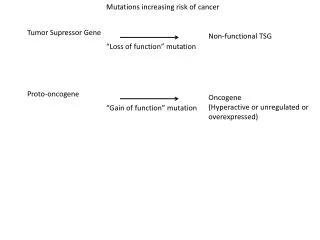
Oncogenes • Oncogenes are mutant forms of the genes for proteins that regulate the Cell Cycle • Oncogenes were originally discovered in tumor-causing viruses and later found to be closely similar to or derived from genes in the animal host cells (proto-oncogenes) • Oncogene generally encode for growth-regulating proteins • During viral infections, the DNA sequence of a protooncogene is sometimes copied or taken by the virus and incorporated into its genome • At some point during the viral infection cycle, the gene can become defective by truncation or mutation. • When this viral oncogene is expressed in its host cell during a subsequent infection, the abnormal protein product interferes with normal regulation of cell growth, sometimes resulting in a tumor.
Oncogenes encode defective signaling proteins. • By continually giving the signal for cell division, they lead to tumor formation. • Oncogenes are genetically dominant and may encode defective growth factors, receptors, G proteins, protein kinases, or nuclear regulators of transcription.
Oncogenes • Oncogenes have been shown many times to be associated with cancer and uncontrolled cellular growth. This growth can lead to tumors. • Two types of tumors exist. • Malignant tumors can induce secondary tumors by the release of cells that can lodge and begin growing in another location of the body. • Benign tumors are cells that remain in the initial location.
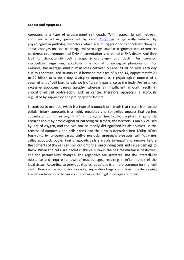
Levels of Cyclin-Dependent Protein Kinase Oscillates • Variations in the activities of specific CDKs during the cell cycle in animals. • Cyclin E–CDK2 activity peaks near the G1 phase–S phase boundary, when the active enzyme triggers synthesis of enzymes required for DNA synthesis. • Cyclin A–CDK2 activity rises during the S and G2 phases, then drops sharply in the M phase, as cyclin B–CDK1 peaks.
pRb functions in most, perhaps all, cell types to regulate cell division in response to a variety of stimuli. Unphosphorylated pRb binds the transcription factor E2F; while bound to pRb, E2F cannot promote transcription of a group of genes necessary for DNA synthesis In this state, the cell cycle cannot proceed from the G1 to the S phase, the step that commits a cell to mitosis and cell division. The pRb-E2F blocking mechanism is relieved when pRb is phosphorylated by cyclin E–CDK2, which occurs in response to a signal for cell division to proceed. When the protein kinases ATM and ATR detect damage
Seven types of proteins that participate in controlling cell growth and proliferation. • Cancer can result from expression of mutant forms of these proteins. • Mutations change the structure or expression of proteins that normally promote cell growth generally give rise to dominantly active oncogenes. • extracellular signaling molecules (I), • Signal receptors (II), • signal-transduction proteins (III), • Transcription factors (IV) • Encoded by tumor-suppressor genes • Cellcycle control proteins (VI) that function to restrain cell proliferation • DNA-repair proteins (VII) are • . Mutations in these genes act recessively, greatly increasing the probability that the mutant cells will become tumor cells or that mutations will occur in other classes. • Tumor suppressors that promote apoptosis and include oncoproteins that promote cell survival • Apoptotic proteins (V). • Virusencoded proteins that activate signal receptors (Ia) also can induce cancer.
Tumor suppressor genes • Tumor suppressor genes encode proteins that normally restrain cell division. Mutation in one or more of these genes can lead to tumor formation. Mutations in tumor supressor genes genes are genetically recessive i.e. tumors form only if both chromosomes of a pair contain a defective gene. • In a person who inherits one correct copy and one defective copy, every cell has one defective copy of the gene. If any one of those 1012 somatic • cells undergoes mutation in the one good copy, a tumor may grow from that doubly mutant cell. • Mutations in both copies of the genes for pRb, p53, or p21 yield cells in which the normal restraint on cell division is lost and a tumor forms. • Mutations in both copies of the genes for pRb, p53, or p21 yield cells in which the normal restraint on cell division is lost and a tumor forms.
Retinoblastoma is a cancer of the retina that occurs in children who have two defective Rb alleles. Very young children who develop retinoblastoma commonly have multiple tumors in both eyes. • Each tumor is derived from a single retinal cell that has undergone a mutation in its one good copy of the Rb gene. (A fetus with two mutant alleles in every cell is nonviable.) • Retinoblastoma patients also have a high incidence of cancers of the lung, prostate, and breast. • p53: Mutations in the gene for p53 also cause tumors; in more than 90% of human cutaneous squamous cell carcinomas (skin cancers) and about 50% of all other human cancers, p53 is defective. Those very rare individuals who inherit one defective copy of p53commonly have the Li-Fraumeni cancer syndrome, in which multiple cancers (of the breast, brain, bone, blood, lung, and skin) occur at high frequency and at an early age.
Mutations in oncogenes and tumor suppressor genes do not have an all-or-none effect. In some cancers, perhaps in all, the progression from a normal cell to a malignant tumor requires an accumulation of mutations (sometimes over several decades), none of which, alone, is responsible for the end effect. For example, the development of colorectal cancer has several recognizable stages, each associated with a mutation • Example • If a normal epithelial cell in the colon undergoes mutation of both copies of the tumor suppressor gene APC (adenomatous polyposis coli), it begins to divide faster than normal and produces a clone of itself, a benign polyp (early adenoma). For reasons not yet known, the APC mutation results in chromosomal instability; whole regions of a chromosome are lost or re-arranged during cell division. This instability can lead to another mutation, commonly in ras, that converts the clone into an intermediate adenoma. A third mutation (probably in the tumor suppressor gene DCC) leads to a late adenoma. Only when both copies of p53 become defective does this cell mass become a carcinoma, a malignant, life-threatening cancer. • The full sequence therefore requires at least seven genetic “hits”: • two on each of three tumor suppressor genes (APC, DCC, and p53) and one on the protooncogeneras.
Many cells can precisely control the time of their own death by the process of programmed cell death, or apoptosis (app-a-toe-sis; from the Greek for “dropping off,” as in leaves dropping in the fall). The regulatory mechanisms that trigger apoptosis involve some of the same proteins that regulate the cell cycle. The signal for suicide often comes from outside, through a surface receptor. Tumor necrosis factor (TNF), produced by cells of the immune system, interacts with cells through specific TNF receptors. These receptors have TNF-binding sites on the outer face of the plasma membrane and a “death domain” of about 80 amino acid residues that passes the self-destruct signal through the membrane to cytosolic proteins such as TRADD (TNF receptor-associated death domain) Another receptor, Fas, has a similar death domain that allows it to interact with the cytosolic protein FADD (Fas-associated death domain), which activates a cytosolic protease called caspase 8. This enzyme belongs to a family of proteases that participate in apoptosis; all are synthesized as inactive proenzymes, all have a critical Cys residue at the active site, and all hydrolyze their target proteins on the carboxyl-terminal side of specific Asp residues (hence the name caspase).
When caspase 8, an “initiator” caspase, is activated by an apoptotic signal carried through FADD, it further self-activates by cleaving its own proenzyme form. Mitochondria are one target of active caspase 8. The protease causes the release of certain proteins contained between the inner and outer mitochondrial membranes: cytochrome c and several “effector” caspases. Cytochrome cbinds to the proenzyme form of the effector enzyme caspase 9 and stimulates its proteolytic activation. The activated caspase 9 in turn catalyzes wholesale destruction of cellular proteins, a major cause of apoptotic cell death. One specific target of caspase action is a caspase-activated deoxyribonuclease . In apoptosis, the monomeric products of protein and DNA degradation (amino acids and nucleotides) are released in a controlled process that allows them to be taken up and reused by neighboring cells. Apoptosis thus allows the organism to eliminate a cell without wasting its components.
Initial events of apoptosis. Receptors in the plasma membrane (Fas, TNF-R1) receive signals from outside the cell (the Fas ligand or tumor necrosis factor (TNF), respectively). Activated receptors foster interaction between the “death domain” (an 80 amino acid sequence) in Fas or TNF-R1 and a similar death domain in the cytosolic proteins FADD or TRADD. FADD activates a cytosolic protease, caspase 8, that proteolytically activates other cellular proteases. TRADD also activates proteases. The resulting proteolysis is a primary factor in cell death
■ All cells require trophic factors to prevent apoptosis andthus survive. In the absence of these factors, cells commit suicide. ■ Genetic studies in C. elegans defined an evolutionarily conserved apoptotic pathway with three major components: regulatory proteins, adapter proteins, and effector proteases called caspases in vertebrates (see Figure 22-31). ■ Once activated, apoptotic proteases cleave specific intracellular substrates leading to the demise of a cell. Adapter proteins (e.g., Apaf-1), which bind both regulatory proteins and caspases, are required for caspase activation. ■ Pro-apoptotic regulator proteins (e.g., Bax, Bad) promote caspase activation, and anti-apoptotic regulators (e.g., Bcl-2) suppress activation. Direct interactions between pro-apoptotic and anti-apoptotic proteins lead to cell death in the absence of trophic factors. Binding of extracellular trophic factors can trigger changes in these interactions, resulting in cell survival ■ The Bcl-2 family contains both pro-apoptotic and antiapoptotic proteins; all are single-pass transmembrane proteins and engage in protein-protein interactions. Bcl-2 molecules can control the release of cytochrome c from mitochondria, triggering cell death. ■ Binding of extracellular death signals, such as tumor necrosis factor and Fas ligand, to their receptors activates an associated protein (FADD) that in turn triggers the caspase cascade leading to cell murder.
Specific examples of cancers • smokers who work with asbestos are more likely to develop lung cancer than smokers who don't work with asbestos because the two carcinogens both play roles in cancer development. • if you've inherited a genetic mutation that predisposes you to cancer, you may be more likely than other people to develop cancer when exposed to a certain cancer-causing substance