Download
1 / 37
370 likes | 389 Views
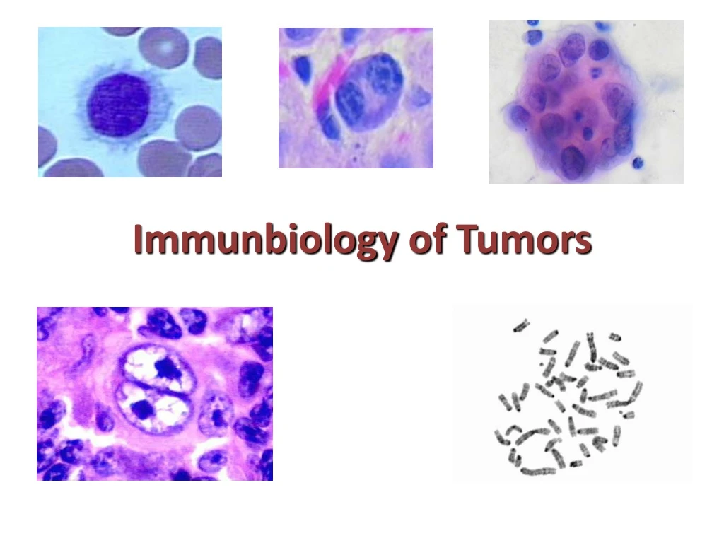
Explore the immunobiology of tumors, including tumor cell formation, oncogenes, neoplastic transformation, tumor antigens, immune responses, and tumor markers. Learn about the host immune response in both experimental and clinical settings.

E N D
Tumor cells are considered as “parasites” of the body • They are formed also in healthy subjects but anti-tumor control mechanism eliminate them • Tumor cells escape control with - fast proliferation - mutations - high diversity (“mini evolution”)
Inducers of malignant transformation of cells • chemical carcinogens e.g. tar • physical carcinogens e.g. UV or X-ray • viral - both DNA or RNA viruses
Oncogens • gene coding a protein which can induce malignant transformation - viralorv-onc(they are only exons) • proto-oncogens - are present and function in physiologicallyintact cells - cellular or c-onc(they are exons or introns) • these genes arewell-conservedstructures • their main function is: to modulate proliferationorapoptosis
Neoplastic transformation • genetic alterations • environmental factors • expression of new, non-self cell surface antigens • the non-self antigens are detected by the immune system
Potentialsources of tumor antigens CategoryExample Oncogeneproductmuationin RAS codon 12 (pancr. cc.) bcr/abl protein (chr. myeloidleukemia) Embryonicproteins MAGE family (melanoma, breastcc.) ViralproteinsEpstein-Barrvirus (Hodgkin’slymph.) Hepatitis B (hepaticellularcc.) Tissuespec. antigenTyrosinase (melanoma) Mutant tu. suppressorprot. p53 (severalcc.) IdiotypicepitopesT-cell receptor idiotypes (T-celllymph.)
Formation of Tumor Specific Antigens effect of mutagen mRNA protein MHC changed peptide
Tumor antigens • Tumor specific antigens (TSA) expressed by tumor cells only • Tumor associated antigens (TAA) expressed by tumor cells and some normal cells, too
Characterization of TSA (1) • expression of TSA is induced by chemical carcinogens or X-rays. • somatic mutations result TSA • each carcinogen can induce unique or specific class of TSA
Characterization of TSA (2) • T-cell receptors recognize peptides bound to the antigen - binding to the binding cleft of MHC molecules • TSA that evoke cytotoxic T cell response are derived from peptides uniquely synthesized by tumor cells andexpressed on the surface of MHC I.
CD8+ T cell Normal cell Normal cytoplasmic protein Peptide fragment MHC I antigen CD8+ T cell CD8+ T cell
Responseselicitedby tumor cells Antigen-specific effects on the tumor cell Escapemechanisms Immunologictolerance Apoptosis of tumor cells METASTASIS,DEATH SURVIVAL
Antigen-specific effect of Tc cells MHC I. TCRa Tumor cell Tc TCRb CD8 TNFb FasL Fas APOPTOSIS APOPTOSIS
Mechanisms by which TSAs are derived • Peptides without structural motives for binding to MHC molecules of the host • Mutation in gene encoding converting proteins that convert the peptide to a form binding to MHC and displayed az TSA on cell surface
40% melanomas expressMAGE-1 (melanomaantigen-encodinggene) 20% breast ca. 30% lung small cell ca. MAGE-1 - embryonal protein, expression is de-repressed in tumors - presentalso in some normal cells e.g. testis
TAA • Oncofetal antigens • Differentialtion specific antigens (DSA)
Oncofetal antigens • normally expressed during a specific phase of enbryogenesis • they are practically in mature, differentiated tuissues • they are not immunogenic • they do not possess functional role in tumor immunity • Their significance: they are diagnostic and prognostic markers • serum cc. correlates with tumormass, level of differentiationand response to therapy
Ideal tumor markers • specific for the typeof the tumor • released onlyin response to tumor • results proportional to tumormass • quantitatively reflects to tumorresponse • elevated even with lowtumor burden
Carcinoembryonic antigen (CEA) • discovered in extracts of adenocc. of colon • a group of heterogeneous glycoproteins - M.W. 200 kD • normally present in embryonic and fetal digestive tissues • detected by RIA or immunoenzymatic technique • elevated (over 5 ng/ml)in gastrointestinal, breast, pancreas, lung tu. and alcoholic cirrhosis, inflammations
Alpha-fetoprotein (AFP) • increased in hepatocellular cc. and malignant teratomas • serum levels are increased in metastatic tumors in liver and acute hepatitis
Prognostic roles of tumor markers † † † 1000 Se AFP ng/ml 100 chemotherapy - no response chemotherapy - response surgery with regrowth or metastasis surgery
In experimental model TSA assessed by: • ability to resist a live tumor implant following the immunization with tumor cells • ability to resist when the model is infused with sensitized T cells from a syngeneic donor • in vitro demonstration of tumor cell destruction by cytotoxic T cells gained from a tumor immunized animal
Host immune response to tumorExperimental • Colony inhibition of tumorsby sensitized lymphocytes • Tumor extracts induce lymphocyte blast tratnsformation • Lymphocyte-enhanced cytotoxicity • Macrophage-enhanced phagocytosis
Host immune response to tumorsClinical • Spontaneous regression • Regression of tu. - sublethal doses of chemotherapy • Regression of metastasis - resection of primary tumor • Mononuclear cell infiltration • High incidence of tu. after clinical immunospureassion • High incidence of tu in immunodeficiency • Increased incidence of tu. in aging
Cellular effectors that mediate immunity (1) • Cytotoxic T-Ly - protective role against virus- associated neoplasms (e.g. EBV) • NK-Ly - capable to destroy tumor cells w\o prior sensitization • Only tu. cells w\o MHC are lysed: NK-Ly recognize carbohydrate components of the membrane by NKR-P1 rec. BUT Ly49 rec. recognize MHC I. and blocks NK-Ly activity (This is the first-line of defense.) • Activation with IL-2: NK Ly lyse a variety of tu. T Ly Complementary anti-tumor mechanisms NK Ly
Cellular effectors that mediate immunity (2) • Macrophages (activated) possess selective cytotoxicity against tumor cells T Ly NK Ly collaborate in anti-tumor reactivity Macrophages (e.g. INF-g secreted by T and NK Ly activator of macrophages. Production of reactive oxygen, or secretioon of TNF-a) • Humoral mechanisms: activation of complement induction of ADCC by NK Ly
Cellular effectors of antitumor immunity IL-2 CTL NK Ly Peptide TC-rec. NK-rec. MHC I Antigen IgG INF-g TNF-a Fc-rec. Macrophage NK Ly
Effector Mechanisms in Tumor Immunity Humoral: • opsonisationand phagocytosis • complementmediated lysis • loss of cell adhesion(antibody dependent) Cellular: • T Ly • Antibody-dependent cytotoxicity • NK Ly • Lymphokine-activated killer (LAK) cells • Macrophages (also activated by lymphokines)
Immuno Surveillance • In5% with congenital immunodeficiencies develop cancer (200x rate) • Inimmunsuppressed patients (80x rate) • AIDS, lymphomas, chronic infectious mononucleosis or malignant lymphomas
Escape of Tumors • Continuousshedding of tumor antigens - solubile antigens Reults: - impossible to detect selectively these cells - only NK-Ly can detect these cells, BUTthey are not present everywhere
KAR KIR + Effects of NK on the target cells Killer inhibitory receptor = KIR; Killer activator receptor = KAR target MHC I KAR KIR + - NK-Ly NO apoptosis Apoptosis
More Escape Mechanisms from Immuno Surveillance • selective outgrowth of antigen negative variants • loss or reduced expression of HLA antigens • no costimulation- no sensitization- anergic T Ly - apoptosis of T Ly • expression of local inhibitorymolecules(TNF- borFas ligand • immunosuppression (chemicals, radiation, TGF-b)
Immunologicactivation and tolerance Viralinfection Cancer V Dangersignals (GM-CSF, TNF-a) Antigen uptake by activated APC T cell V V Costimulatory signal (B7, Il-12) T cell T cell Tumor-specific T cellsbecome tolerant Virus-specific T cellsbecome activated
Failure of Immune Response to Tumor • Some tumors arise in areas not accessible to the effector cells (e.g. eye, CNS) • Antigenic modulation - antigensof tumor cells may undergo several changes • Blocking factors - immune complexes or cytophilic antibodies can mask tumor antigens or prevent binding by effector cells or lytic antibodies
Prospects for Immunotherapy to generate antitumor responses (1)1890 WiliamColey: bacterialextractstoactivategeneralimmunity • Tumor cell vaccines - there areT cells directed against the tumor - their number could be increased with autologous or allogeneictumor cells (the replication is prevented by irradiation) • Immunization with tumor-specific peptides - only for tumors for whichTAAhave been cloned and peptides synthesised • Cytokine therapy - increasingthe levels of some cytokines (IL-2, IFN, GM-CSF, IL-7, IL-12) Problems:short life-time, toxicity, non-specific cytokines.
Prospects for Immunotherapy to generate antitumor responses (2) • Monoclonal antibodies - used to deliverimmunotoxins. radioisotopes, drugs. Bivalent antibodies recognize both T cells and TAA - they guide T cell to the tumor. • Gene therapy - combines the concepts of “tumor cell vaccines” and “cytokine therapy” by expressing genes coding for cytokines, costimulatory molecules or MHC. • Adoptive immunotherapy - with antitumor cells, tumor infiltrating lymphocytes (TIL) or lymphokine-activated killer (LAK) cells. Problems: growing the large number of cells required, loss of antigene specificity for T cells, altered homing pattern