Download
1 / 70
710 likes | 937 Views
Hypersensitivity reactions. Prof . Mohamed Osman Gad El Rab. College of Medicine & KKUH. Introduction: Immune reactions leading to pathological tissue damage. - Occur as :

E N D
Hypersensitivityreactions. Prof . Mohamed Osman Gad El Rab. College of Medicine & KKUH.
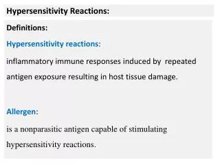
Introduction: Immune reactions leading to pathological tissue damage. - Occur as : 1.Secondary heightened (increased) immune responses . OR 2.Secndary inappropriate (abnormal ) immune responses.
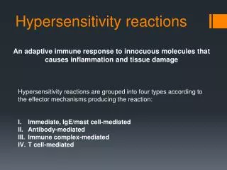
Four major categories according toCoombs and Gell classification : • Type I : Immediate H/S. • Type II : Cytotoxic H/S. • Type III : Immune – complex H/S. • Type IV : Delayed H/S.
Types I , II and III : are mediated by antibodies . • Type IV : Is generated by cell-mediated immune responses.
Hypersensitivity reactions differ inthe rate at which they occur : • Type I : Can occur within minutes afterexposure to antigen. • Type II and III : time course , (4-8) hours todays . • Type IV : require 2 - 4 days.
Hypersensitivity reactions : - can occur as isolated reactions, OR - more than one reaction can occur in the same patient. e.g. : Type I and Type III.
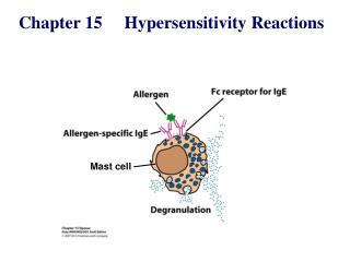
Type I Hypersensitivity. • Also termed : *Immediate H/S ( can literally occur within minutes to hours ). * Anaphylactic reactions . OR * Allergic reactions.
Features : • Antibody isotype : IgE . - Cellular components: Mast cells , basophiles & eosinophils. - Antigens : termed allergens ( antigens with low molecular weight & highly soluble.
Type I H/S : * Evolved as a defense against parasitic infections. * However , many reactions in some predisposed individuals are directed towards harmless molecules (allergens) and these are said to be : “atopic.”
Atopy. • Occur in certain genetically predisposed individuals . • They comprise approx. 15 – 20 % of the population . • Atopy tend to run in families .
The likelihood to generate a strong IgE response is determined by : - genetic factors . - environmental factors. these factors depend on exposure to allergens of diverse nature ( pollens , foods , drugs fungal spores , bee - sting venoms , house dust mites and animal dander.
Type I Reaction occur in 2 phases: • Phase I : - Sensitization phase . Allergen enter tissues , induce an immune response . B – cells transform to plasma cells & produce IgE. - IgE bind to receptors on Mast cells and basophiles ( FcЄRI - high affinity receptors). individuals become : “ Sensitized . “
Phase II : Challenge phase . -Subsequent encounter with same allergen cross – link IgE on Mast cells . -This generate an intracellular signal that prompts the Mast cells to: “ Degranulate”
Development of Allergy Requires Sensitization , Re - exposure & Degranulation Naive Mast Cell Sensitized Mast Cell Degranulated Mast Cell
Degranulation : • The release of a wide variety of mediators of inflammation . • These exert effects on surrounding target tissues. • There are 2 types : 1. primary mediators. 2. secondary mediators.
Primary mediators. • 1. Histamine ,heparin. • 2. Serotonin . • 3. Eosinophil chemotactic factor (ECF). • 4. Neutrophil chemotactic factor (NCF ). • 5. Proteases .
Secondary mediators : • 1. Platelet activating factor . • 2. Leukotriens ( slow reacting substance of anaphylaxis). • 3. Prostaglandins . • 4. Bradykinin. • 5. Cytokines .( IL-1,TNF-a , IL-2 , 3, 4, 5, 6, )
Some effects of mediators : • Amines & active peptides - Histamine ( increase vascular permeab.). (constriction of vasc. smooth muscle). - Heparin ( counters coagulation ). • Cytokines & chemokines : -IL – 5 (eosinophils). -IL – 8 (neutrophils). - IL – 4. ( IgE) • Enzymes – Elastase : ( reconstruction of connective tissues).
Some effects of secondary mediators : Prostaglandins :vasodilatation, contraction of pulmonary smooth muscle . Leukotrienes :increased vascular permeab. contraction of smooth muscle. Platelet-activating factor: platelet aggregation, contraction of smooth muscle .
Eosinophils:- • Major basic protein (MBP) activate mast cells and basophiles. • Eosinophil Cationic protein (ECP) toxic to parasites. • Mast cells and Basophiles interact with Eosinophils (can bind IgE). • Eosinophils release:- - Enzymes. - Cytokines. - Chemokines. • Therefore, contribute to the inflammatory reaction.
Elevated IgE. • Blood eosinophilia. are clinical signs of Type I reactions.
Type 1 reactions result in : * Vasodilatation and increased capillary permeability . * Edema. * Vasoconstriction ( arteries and arterioles ) * Bronchoconstriction. * Increased mucus secretion.
Symptoms of an allergic (Type I) reaction are determined by location of allergens:- • Inhaled allergenswhen deposit in nasopharyngeal and bronchial tissues result in : - Allergic rhinitis. - Allergic asthma. • Ingested allergens : food allergy (G.I.T symptoms)
Bee sting allergens Injected into the blood. Systemic inflammation. Anaphylactic shock. (life - threatening). • Anaphylactoid reactions:- are non - IgE mediated. may result from contrast media or local anesthetics.
Diagnosis:- 1. Skin prick test (SPT). 2. Intradermal test. 3. Specific IgE measurement (RAST). 4. Challenge test ( Nasal , Bronchial). 4. Elimination / Provocation test (Food allergy).
Type II Hypersensitivity.(Cytotoxic H/S). • Features:- - IgG. - Antigens ( Bound to cell membranes or extra cellular matrix). - Self - antigens. - Exogenous antigens. (microbial ) - Complement activation (Invariable).
Mechanisms of tissue damage in type 11:- • IgG (from blood) fix to bound antigen. - Activate complement. - Complement generate chemotactic agents (C5a). - Attract neutrophils and other inflammatory cells.
Neutrophils bind to target through:- A. Complement receptors -(immune -adherence). B. Antibody receptors - (Opsonic -adherence). - They secrete their enzymes to the outside (Exocytosis). - They cause direct damage.
Complement - mediated damage (type11):- • Activation of complement C8 , C9. - Membrane attack complex (MAC). - Direct lytic damage on target tissues.
Clinical Examples ( type 11):- 1, Incompatible blood transfusion (ABO). -massive intravascular hemolysis of RBC. -Immediate reactions-IgM mediated. -Delayed reactions-(2-6 days ),IgG mediated. 2. Hemolytic disease of the new -born (HDN). 3. Drug reactions (Drugs bind to R.B.C , W.B.C , platelets). - Lead to:- - Hemolytic anemia. - Thrombocytopenia. - Leucopenia.
4. Autoimmune diseases (Self-antigens). 5. Graft rejection (Hyper - acute). - Preformed antibodies in the recipient . Diagnosis:- - Detection of antibodies and antigens by immunofluorecence. (Biopsy).
Type III Hypersensitvity(Immune - complex H/S ):- • Features:- - IgG or IgM. - Soluble antigens. - Immune – Complex formation. - Complement activation. (invariable).
Mechanism of tissue damage (type 111):- • Immune - Complexes are continuously forming. - Depend on the nature and conc. of antigens and antibodies. - As long as they are not :- - Extremely large. - Numerous. they are readily cleared.
Immune complex clearance : 1. The mononuclear- Phagocyte system. - Macrophages and dendritic cells degrade particles and debris. 2. Erythrocytes bind complexes via FcR1 receptors and release them in the liver.
When size and quantity over whelm the normal clearance mechanism:- 1. Complexes accumulate and deposit in blood vessels and tissues. 2. They activate complement. Therefore : induce immune - complex disease.
Mechanism of tissue damage (type 111): Complexes deposit in blood vessels and tissues and result in vasculitis , arthritis …et . • Two main types : 1. Complexes with antibody excess , are termed Arthus – type reactions (localized ). 2. Complexes with antigen excess , are termed serum – sickness reactions (systemic).
Sites susceptible to type III H/S: 1. Glomeruli . - high blood flow . - filtration of the blood. - complement receptors . Lead to glomerulonephritis . 2. Blood vessel walls . - complexes deposit on walls of veins and arteries . • Lead to vasculitis .
Type 111,cont. 3. Synovial membrane of joints . - immune- complexes. deposition can damage bone and cartilage . 4. Skin . - a common site for deposition of immune complexes manifest as rashes .
Clinical examples (type 111) : 1. Autoimmune disease ,(self – antigens ). 2. Chronic infections ,(microbial antigens) . 3. Cancer , (tumor antigens). 4. Drug reactions , (chemical haptens ).