
Basic tissues
E N D
Presentation Transcript
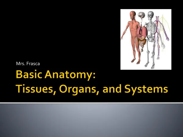
Basic tissues • Epithelium • Connective tissues • Muscles • Nervous tissue Jeanne Adiwinata Pawitan Dept. of Histology FMUI Jeanne A Pawitan
Basic tissues • Epithelium: cells, intercellular substance/extracellular matrix – basement membrane • *Connective tissue: intercellular substance↑ • Muscle: muscle cells* = muscle fibers • Nerve*: • Neurons • Neuroglial cells (support, protect neurons) Jeanne A Pawitan
Epithelium Jeanne A Pawitan
Layers Single Simple (Pseudostratified) Multiple Stratified (Transitional) Shape Squamous Cuboidal Columnar (torax) Location Epithelium (and glands) Jeanne A Pawitan
Polarity and cell surface specializations • Morphological domain • Biochemical domain Polarity • Functional domain • Apical domain – free surface – lumen • Basolateral domain • Basal - basal lamina • Lateral – other cells Jeanne A Pawitan
Microvilli Cilia Kinocilia Stereocilia Ion channels Aquaporins Carrier proteins H+- ATPase Glycoproteins Hydrolytic enzymes Apical domain - lumen Jeanne A Pawitan
Basolateral domain • Intercellular interdigitation, basal enfoldings • Junctional specialization • Receptors • Hormones • Neurotransmitters • Na+-K+ ATPase • Ion channels • Sites for constitutive secretions Jeanne A Pawitan
Junctional specialization • Lateral membrane specialization – terminal bars (LM) – junctional complexes (EM) • Occluding junction, tight junction, zonula occludens • Adhering junction • Zonula adherens -zonulae adherentes • Fascia adherens • Macula adherens (desmosome) - hemidesmosome • Gap junction Jeanne A Pawitan
Connective tissue Jeanne A Pawitan
Connective tissues • Mesenchyme (embryo) – mucosa /gelatinosa • Connective tissue proper • Loose (areolar) connective tissue areolar CT • Reticular connective tissue • Adipose tissue • Dense connective tissue • Dense regular connective tissue • Dense regular collagenous connective tissue • Dense regular elastic connective tissue • Dense irregular connective tissue • Specialized connective tissue (cartilage, bone, blood) Jeanne A Pawitan
Cells of connective tissue • Mesenchymal cells - mesenchyme • Fibroblasts/fibrocytes • Histiocytes/macrophages • Mast cells • Pericytes • Adipose cells • Reticular cells – reticular CT • Transient cells: plasma cells, leucocytes, macrophages Jeanne A Pawitan
Intercellular substance/ extracellular matrix • Ground/amorphous substance (gel like)– tissue fluid • Glycosaminoglycans (GAG) (hyaluronic acid, heparin, sulfated GAG) • Proteoglycans ( certain GAG – Protein) • Adhesive glycoproteins (fibronectin, laminin, entactin, tenascin, chondronectin, osteonectin) • Fibers • Collagen type I – VII • Reticular (type III collagen) • Elastic fibers Jeanne A Pawitan
Basement membrane • Basal lamina • Lamina lucida • laminin, entactin • Transmembrane protein: integrin, dystroglycan • Lamina densa (perlacan-type IV collagen meshwork-perlacan, fibronectin) • Lamina reticularis: type I, III collagen Jeanne A Pawitan
Muscles Jeanne A Pawitan
Muscles • Striated muscle – characteristic striation – specific arrangement of myofilament (contractile protein in a myofibril: myosin, actin) • parallel arrays regularly repeated arrangement • interdigitating in myofibril • skeletal muscle • cardiac muscle • Smooth muscle • Myoepithelial cells –glandular unit in glands • Myofibroblast – wound contraction, tooth eruption Jeanne A Pawitan
Unique terms in muscle • Sarcolemma = cell membrane • Sarcoplasm = cytoplasm • Sarcoplasmic reticulum = smooth surfaced endoplasmic reticulum (SER) • Sarcosome = mitochondria • muscle fiber = muscle cell Jeanne A Pawitan
Smooth muscle • Location: wall of hollow organ - lumen • Digestive system • Reproductive system • Urinary system • Respiratory system • Ducts - large (glands) • Dermis (skin) • Function: • contraction • Protein synthesis extra cell: collagen, elastin, glycosaminoglycans, proteoglycans, growth factors Jeanne A Pawitan
Smooth muscle • contraction - involuntary – controlled by • Autonomic nervous system (sympathetics, parasympathetics) • Hormone: bradykinin • Local physiological condition • Every fiber – lined by external lamina (outside sarcolemma) • reticular fiber - embedded in external lamina – surrounding each muscle cell – function: control and unify the strength of muscle fiber contraction Jeanne A Pawitan
Smooth muscle fiber • Shape : fusiform, elongated –Ø 5-6 μm, length -0.2 mm • Surrounded by sarcolemma (cell membrane) • Nucleus - center – oval – 1-2 nucleolei – at contraction nucleus bottle opener • HE: cytoplasm – homogenous – acidophilic • Iron hematoxylin – in sarcoplasm: • Dense body – attached at the inside of cell membrane • Longitudinal lines groups of myofilaments Jeanne A Pawitan
Smooth muscle fiber • Form sheets/layers – usually 2 layers – perpendicular • fibers –closely packed – thin part >< thick part • Cross section – fiber diameter ≠ – section - nucleus (+)/(-) • Individual cell Jeanne A Pawitan
Innervation – smooth muscle fiber • Neuromuscular junction usually en passant type • Bulging of axon • Containing synaptic vesicles • Nor epinephrine – sympathetic • Acetylcholine – parasympathetic • Type • multi unit –individual innervation (iris, vas deferens • Intermediate (mixed 30-60% - individual) • Visceral – some fibers – 1 neuromuscular junction – gap junction/nexus (uterus, GI tract) – other influencing factor • Humoral factor : oxytocin • Micro environmental factor : stretching (intestines) Jeanne A Pawitan
Cardiac muscle • Development: Myoepicardial mantle in splanchnic mesenchyme epicardium + myocardium • Cardiac muscle • Cardiac muscle fiber–branching-anastomosing network – arranged in layers (laminae) • Between fibers – connective tissue • Connective tissue between laminae – contains: • Blood vessels capillaries network –in connective tissue between each fiber • Nerves • conducting system – in heart Jeanne A Pawitan
Cardiac muscle fiber • Striated, branching • Length 15-80 μm • Nucleus 1 (occasionally 2) – center – big –oval • Between cells (at the end) - intercalated disk-consist of: • Transverse portion: facia adherens ( Z disk), desmosome • Lateral portion: gap junction (also found in regions - side by side contact – long.) Jeanne A Pawitan
Sarcoplasm contains • Myofibrils Ø-1-2 μm –longitudinal –regularly arranged striated (longitudinal section): • A band – (anisotropic on polarized light dark) • I band– (isotropic on polarized light) • EM: Sarcoplasmic reticulum = SER • Network around myofibril • Function: intracellular Ca storage (< skeletal muscle) • Small terminals (terminal cisternae -) • The rest = longitudinal sarcotubule + branches • Contraction needs active transport of Ca from extra cell • membrane contains junctional feet (= voltage gated Ca release channels) Jeanne A Pawitan
Sarcoplasm contains • Organels • Mitochondria >>> - ½ volume – cardiac muscle • Myoglobin O2 transport • Glycogen particle energy source for contraction ( with lipid) Jeanne A Pawitan
Cardiac muscle fiber – T tubule • EM: Sarcolemma – tubular invagination – transverse – branching - anastomosing T tubule (transverse tubule) • Intertwine among myofibril • Location: around Z line – 1/sarcomere • Open to extra cellular compartment -lined by external lamina • Ø 2.5 X T tubule in skeletal muscle • Function: facilitate conduction – depol. wave -along sarcolemma • Associated with sarcoplasmic reticulum: T tubule + small terminals dyad Jeanne A Pawitan
Myofibrils • A band (dark) – H band (light) – M line • I band (light) – Z line (Z disk, dark) • Sarcomere = myofibril region between 2 Z line = 1 contractile unit • Contraction: • I band - shortened • H band - disappeared • A band - constant • Z line - approaching each other Jeanne A Pawitan
Cardiac muscle fiber-atrium • Ø < fiber in ventricle myoendocrine cells • Contains granules - cardiodilatin (CDD)/Atrial Natriuretic (Poly)peptide (ANP) – esp. in right atrium • ANP – regulation of fluid –electrolyte balance • Efferent arteriole constriction diuresis, sodium excretion↑ • Sodium, water resorption in kidneys (renal tubules)↓ blood pressure ↓ - • Vasodilatation • inhibit the release of arginine-vasopressin (posterior hypophysis), and aldosterone (adrenal cortex) Jeanne A Pawitan
Skeletal muscle • investments • Epimysium – muscle – irregular dense collagenous connective tissue • Perimysium – fascicles (bundles) - < dense • Endomysium – muscle fiber (cell) - reticular fiber, basal lamina / external lamina • cell • Skeletal muscle fibers – striated – parallel • Satellite cells Jeanne A Pawitan
Skeletal muscle cells/fibers • Development: myoblast – parallel end –end fused myotube – formed myofibril skeletal muscle fiber • Shape: long, tubular – Ø 10-100μm • Between fibers: endomysium + continuous capillaries – parallel to fiber • Skeletal muscle fiber – non branching -striated - multinucleated • Nuclei – periphery – in a row – inside cell membrane (sarcolemma) • EM: Sarcolemma invaginate T tubule (transverse tubule) Jeanne A Pawitan
Sarcoplasm: contains • A lot of myofibril Ø-1-2 μm –longitudinal – along the fiber – regularly arranged striation in longitudinal section: • A band – (anisotropic on polarized light dark) • I band– (isotropic on polarized light light) • EM: Sarcoplasmic reticulum = smooth surface endoplasmic reticulum • Form a network around myofibril • Function: intracellular Ca storage (esc. terminal cysterna) • A-I junction – dilated terminal cysterna • The membrane has junctional feet (= voltage gated Ca release channels) • The rest = longitudinal sarcotubules + their branches Jeanne A Pawitan
T tubule • Tubular invagination –transversal – branching-anastomosing – intertwine among myofibril • Location: transversal plane - A-I junction 2 in each sarcomere • To extra cell - open • Associated with sarcoplasmic reticulum • Function: facilitate conduction of depol wave along sarcolemma • T tubule + 2 terminal cysternae triad • function: spreading of depol wave – fast – from sarcolemma – trough out the cell – to terminal cysterna Jeanne A Pawitan
Myofibril • A band (dark) – H band – pale –M line • I band (light) –Z line, Z disk – thin, dark • Sarcomere = myofibril area between 2 Z line = contractile unit of skeletal muscle fiber • Contraction: • I band shortened • H band disappeared • A band remain unaltered • Z line – move closer together Jeanne A Pawitan
Satellite cells • Satellite cells = regenerative cells • Small cell – location: shallow depression – muscle cell’s surface • External lamina – shared with muscle cell → inside external lamina • Nucleus-one - > dense - > coarse Jeanne A Pawitan
Myotendinuous junction • Connective tissue–muscle – tendon – continuous • Cells tapered – fluted (deeply folded) • Collagen fibers-tendon – penetrate • Infoldings • Endomysium – continuous – reticular fibers Jeanne A Pawitan
Innervation of skeletal muscle • Motor nerve fibers (α motor neurons –axon-myelinated) branching – unmyelinated (Schwann cells +) – terminal dilated - myoneural junction (motor end plate) • Axon terminal • Synaptic cleft • Muscle cell membrane • Sensory nerve fibers • muscle spindles – among muscle cells –stretch Rx • Golgi tendon organ (neurotendinuous spindle)– inhibitory feedback - α motor neurons - relax Jeanne A Pawitan
Regeneration - muscles • Skeletal muscle – regeneration (+) • Skeletal muscle fiber ≠ mitosis • Satellite cells • Injury: satellite cells - mitosis – hyperplasia • At muscle building: satellite cell – muscle fiber -fusion mass increase hypertrophy • Cardiac muscle - ≠ regeneration, fibroblast – connective tissue (scar) • Smooth muscle – regeneration (+) Jeanne A Pawitan
Regeneration – smooth muscle • Injury formation of new fibers • Mitosis – smooth muscle fiber (GI tr., urin. tr.) • Differentiation - pericyte – blood vessel • Uterus –pregnant – smooth muscle • hyperplasia - mitosis • hypertrophy Jeanne A Pawitan
Nervous tissue Jeanne A Pawitan
Neurons • Cell body (soma, perikaryon) • Dendrite (multiple) - branching • Axon (single) – long - branching • Axon Hillock • Axon terminals, end bulbs, terminal boutons → synapse Jeanne A Pawitan
Specialized cells Jeanne A Pawitan
Specialized cells • Protein secreting cells • Steroid secreting cells • Neuroendocrine cells • Cells in organs → other modules Jeanne A Pawitan
Protein secreting cells • Ex.: chief cells (stomach), acinar cells (pancreas) • Shape: columnar, truncated pyramid, etc • Basal cytoplasm - basophilic • Nucleus • Rich in RER, polysome • Apical cytoplasm - acidophilic • Secretory granules – proenzyme (zymogen) Jeanne A Pawitan
Steroid secreting cells • Ex: spongiocytes (adrenal cortex) • Shape: polyhedral • Cytoplasm • Extensive network of SER • Lipid droplets – appear vacuolated Jeanne A Pawitan
Neuroendocrine cells • Argentaffin, argyrophilic – silver staining • APUD= amine precursor uptake decarboxylation • DNES= diffuse neuroendocrine system • Location: epithelium/glands – systems • Secretory granules –at basal cytoplasm/ near blood vessel Jeanne A Pawitan
Stem cells Jeanne A Pawitan
Stem cells • Fertilization → 2-4 cells → morula (8 cells) →blastocyst • Trophectoderm → placenta • Inner cell mass → embryo ↓ Embryonic stem cells → determined stem cells (connective tissue, blood) → unipotential stem cells → differentiation (precursors of certain cells) → certain cells Jeanne A Pawitan
Stem cells • Totipotent – can give rise to cells of all orders • Pluripotent – can develop in any one of several possible ways, or affect more than one organ/tissue – multipotent • Unipotent – can give rise to cells of one order only Jeanne A Pawitan