Download
1 / 37
410 likes | 740 Views
Thrombosis, Embolism and Infarction. Dr. Raid Jastania. Hemostasis and Thrombosis. Hemostasis is the physiological process of maintaining blood in fluid state and formation of hemostatic plug at site of vessel injury.

E N D
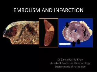
Thrombosis, Embolism and Infarction Dr. Raid Jastania
Hemostasis and Thrombosis • Hemostasis is the physiological process of maintaining blood in fluid state and formation of hemostatic plug at site of vessel injury. • Thrombosis is the pathological process of blood clotting in uninjured vessel or exagurated response to minimal injury. • Components: • Vessel wall • Platelets • Coagulation pathways
Normal Hemostasis: • Vessel injury - brief period of arteriolar vasoconstriction (neurogenic reflex ,endothelin) • Endothelial injury exposes ECM (highly thrombogenic material). • Platelets adhere to endothelial cells and ECM, and are activated. They release their secretory granules. Platelet aggregation occurs forming hemostatic plug (Primary hemostasis) • Tissue factor (produced by endothelium) activates coagulation - formation of thrombin which act of finbrinogen to form fibrin (secondary Hemostsis)
Normal Hemostasis: • The process continues to form the permanent plug formed by polymerized fibrin and platelet aggregates. • At the same time tissue plasminogen activator (t-PA) is formed and it limits hemostatic plug. • Fibrinolysis is also activated to limit heostatic plug to the site of injury.
Thrombosis: • Thrombosis is the pathological process of blood clotting in uninjured vessel or an exaggerated blood clotting response to minimal injury. • Virchow Triad: (factors predisposing thrombosis) • Endothelial injury • Blood stasis or turbulence of blood flow • Blood hypercoagulability
Endothelial Injury • An important factor in arterial thrombosis. • Occurs in myocardial infarction, ulcerated atherosclerosis, trauma, and inflammatory disease of vessels. • Endothelial dysfunction is also a predisposing factor for thrombosis. Eg. Hypertension, bacterial endotoxins, hypercholestrolemia, radiation, cigarette smoking. • Loss of endothelium will expose the ECM and hence activation of platelets and thrombosis.
Blood Stasis and Turbulence of Flow • Turbulence enhances endothelial injury. • Stasis enhances venous thrombosis. • Both result in: • Bringing platelets close to endothelium • Accumulation of clotting factors • Prevent clotting factors inhibitors • Endothelial activation • Example: aortic aneurysm, MI, valve stenosis, rheumatic heart disease, hyperviscosity, sickle cell disease.
Hypercoagulability • It is an alteration in coagulation leading to thrombosis. • Primary: (genetic) • Factor V mutation • Antithrombin III deficiency • Secondary: • Prolonged immobilization • Cancer • Lupus anticoagulant • Nephrotic syndrome • Contraceptive pills • Smoking
Hypercoagulability • Heparin-induced Thrombocytopenia: • When heparin is administered it induces the formation of antibodies that bind platelets and activate them. • Antiphospholipid syndrome (Lupus anticoagulant): • Antibodies to phospholipid (eg. Cardiolipin) • In-vitro: it inhibits coagulation • In-vivo: it induces coagulation
Thrombosis • May develop in the heart, arteries, veins and capillaries. • Arterial thrombi and cardiac thrombi occur at site of endothelial injury or turbulence of flow. • Venous thrombi occur in areas of blood stasis. • Thrombi usually are attached to the underlying vessel wall (mural thrombi) • Arterial thrombi grow back to the heart. • Venous thrombi grow toward the heart.
Thrombosis • Arterial thrombi are firmly attached to the wall and show lines of Zahn (layers of fibrin and platelets alternate with layers of RBC and WBC. • Venous thrombi do no show clear lamination. • In the heart: common causes: MI, dilated cardiomyopathy, arrhythmia, myocarditis, or valvular disease.
Thrombosis • In arteries: common causes: atherosclerosis, and aneurysm. • Arterial thrombi usually occlude the lumen, common in coronary, cerebral, and femoral arteries. • Venous thrombi (phlebothrombosis) are almost always occlusive, Red thrombi, 90% occur in lower extremities.
Fate of thrombus • Propagation (progression) • Embolization • Lysis 4.Organization and recanalization (inflammation and fibrosis)
Venous Thrombosis: • Superficial: eg. Saphenous vein • Local congesion, edema, swelling, pain, tenderness, ischemia, risk of infection • Rarely embolize • Deep Vein Thrombosis: eg. Popliteal, femoral, iliac veins. • Can embolize • There is a lot of collaterals so the congestion and edema are not prominent. • 50% are asymptomatic.
Venous Thrombosis • Blood stasis is common predisposing factor for venous thrombosis. Eg. Heart failure, surgery, trauma, burn, pregnancy, cancer (Trousseau syndrome)
Cardiac and arterial thrombi • MI, valve disease, arrhythmia, atherosclerosis • Possible embolism to brain, kidneys, spleen.
Disseminated Intravascular Coagulation • Sudden widespread fibrin thrombi in the microcirculation • Occurs in pregnancy, and with malignancy. • Leading to circulatory insufficiency: brain , lung, heart, kidneys • Leading to consumption of platelets and clotting factors and risk of bleeding.
Embolism • Detached intravascular solid, liquid or gaseous mass carried by blood to a distant site. • Types: Thrombus 90%, fat, air, cholesterol, tumor, bone marrow, foreign body • Occlusion of vessels and ischemia/infarction
Pulmonary Thromboembolism • 20-25/ 100,000 of hospital patients • 95% coming from DVT (above knee) • may occlude main pulmonary artery (Saddle embolus) • or in small branches of vessels (multiple) • Paradoxical embolus: cardiac embolus passing to the right side through septal defect.
Pulmonary Thromboembolism • 20-25/ 100,000 of hospital patients • 95% coming from DVT (above knee) • may occlude main pulmonary artery (Saddle embolus) • or in small branches of vessels (multiple) • Paradoxical embolus: cardiac embolus passing to the right side through septal defect.
Pulmonary Thromboembolism • 60-80% are asymptomatic • most organize • can lead to cor pulmonale, sudden death. • Result in hemorrhage, and rarely infarction • Obstruction of small vessels lead to small infarctions • Multiple emboli may lead to pulmonary hypertension
Infarction • Ischemic necrosis caused by occlusion of arterial or venous vessles. • Example: MI, cerebral infarction, pulmonary infarct, bowel infract, gangrene • 99% due to thrombosis, mostly arterial • Can be: • Vasospasm • External pressure • Trauma • Twisting of organs eg. Testicular torsion • Edema
Infarction • Venous infarct occurs in organs with single venous outflow. Eg. Testis, ovary • Types: Red infarct, white infarct, septic infarct • Red infarct: • Due to venous occlusion • In loose tissue eg. Lung • Organs with dual circulation • In tissues that have be previously congested • White infarct • Arterial occlusion of solid organs, eg. Heart, kidneys, spleen
Infarction • Infarction is usually wedge shape surrounded by rim of hyperemia • Hemosiderin pigment may accumulate following hemorrhage • Necrosis is of coagulative type (except brain: liquifactive) • Inflammation within few hours • Repair process
Factors influencing development of Infarct • Nature of the blood supply • Dual: lung, liver, hands • End-arterial: spleen, kidneys • Rate of occlusion: • Eg. Atherosclerosis of coronary arteries is gradual slow process
Factors influencing development of Infarct 3. Vulnerability to hypoxia • Neuron: 3-4 minutes • Heart: 20-30 minutes • Fibrous tissue: hours 4. Oxygen content of the blood • Eg. Heart failure patient have low oxygen concentration in blood
Normal Endothelium • Endothelial cells are activated by injury, infection, plasma mediators and cytokines. They have pro-thrombotic and anti-thrombotic functions.
Anti-thrombotic properties: • Anti-platelet effect: • Non activated platelets do not adhere to endothelium. • PGI2, and NO (produced by endothelium) prevent platelet adhesion • Anticoagulant properties: • Heparin-like molecule activate anti-thrombin III • Thrombomodulin binds thrombin which activate protein C (anticoagulant) • Fibrinolytic properties: • Endothelium synthesize t-PA (fibrinolysis)
Pro-thrombotic properties: • Von Willebrand factor: • It enhances binding of platelets to ECM. • Tissue factor • Produced by endothelium, it activates extrinsic clotting pathway • Plasminogen activator inhibitors (PAI)
Normal Platelets • Platelets contain • Alpha-granules: P-selectin, fibrinogen, fibronectin, factor V, factor VIII, PDGF,TGF-alpha. • Delta-granules: ATP, ADP, Ca, histamine, epinephrine
Normal Platelets • On encountering ECM: • 1. Platelets adhere to ECM (collagen) mediated by vWF. • 2. Secrete their granules • 3.Platelets aggregate: forming the primary hemostatic plug which is reversible. With the action of thrombin, platelet contraction occur and the plug becomes irreversible (secondary hemostatic plug)
Normal Platelets • PGI2 inhibits platelet aggregation • Thromboxane A2 (TXA2) enhances platelet aggregation • Aspirin inhibits the synthesis of TXA2
Normal coagulation Cascade • It is series of enzymatic conversions turning inactive proenzymes to active forms. • They lead to formation of thrombin • Thrombin converts fibrinogen to fibrin. • Each reaction needs: enzyme, substrate, cofactor, phospholipid complex, Ca ions • Two pathways: extrinsic and intrinsic: both lead to activation of factor X.
Intrinsic pathway is activated by activation of Hageman factor (factor XII). • Extrinsic pathway is activated by tissue factor. • The process is controlled by anticoagulants: • Antithrombins (eg. Antithrombin III). It is activated by binding to Heparin-like molecule on endothelium. • Protein C and S (vit K dependent) they inactivate factors Va and VIIIa.
Fibrinolytic cascade: • While coagulation occurs • Factor XII or tissue plasminogen activator (t-PA) act on plasminogen to form plasmin. • Plasmin start the process of fibrin lysis an the production of fibrin degradation produces. • The function of plasmin is controlled (opposed) by plasminogen activator inhibitor (PAI) and alph2-antiplasmin.