Download
1 / 34
350 likes | 573 Views
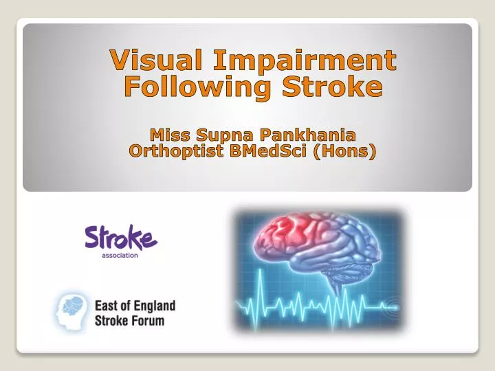
Visual Impairment Following Stroke Miss Supna Pankhania Orthoptist BMedSci ( Hons ). Brain damage due to lack of oxygen and nutrients to the brain as a result of burst blood vessels or a blood clot. ( The World Health Organisation – WHO ). CLINICAL DEFINITION

E N D
Visual Impairment Following Stroke Miss SupnaPankhania OrthoptistBMedSci (Hons)
Brain damage due to lack of oxygen and nutrients to the brain as a result of burst blood vessels or a blood clot. (The World Health Organisation – WHO) • CLINICAL DEFINITION • ‘A rapidly developing episode of focal or global dysfunction, lasting longer than 24 hours or leading to death, and of presumed vascular origin.’ (Macintosh, C; 2003) What is a Stroke?
Leslie Ritter and Bruce Coull, 2011 (The University of Arizona)
85% ischaemic: • Cerebral artery blockage (cerebral thrombosis) Cerebral Infarction • Atheroma build up in intra and extracranial arteries • Embolus – travelling blockage • 15% intracerebral or arachnoid haemorrhage Aetiologies
Cerebral Infarctions 75% 10% } 15%
Most common cause of disability • Third most common cause of death • 150,000 sufferers per year on average • Age 65+ • Effects dependent on area of brain affected • All different in and of variable severity • Effects short and long term Statistics - RNIB
Walking • Speech and or language • Mental processes – cognition • Swallowing • Paralysis • Eyesight Common Effects of Stroke
Visual field loss • Blurring of vision • Reduced vision • Nystagmus • Diplopia • Moving images – oscillopsia • Visual Neglect • Difficulty judging depth and movement • Photosensitivity • Hallucinations • Agnosia – recognition difficulties Visual Effects of Stroke
Ptosis • Anisocoria Visual Effects of Stroke
OCULAR MOTILITY DISORDERS • Diplopia • Gaze palsy • Oscillopsia • Cranial Nerve Palsies – isolated or combined • Supranuclear gaze palsies • Skew deviation • INO Visual Effects of Stroke
Medial rectus, superior rectus, inferior rectus, inferior oblique, levator palpebrae superioris, pupillary sphincter Left Complete 3rd Nerve Palsy
Superior Oblique muscle Right 4th Nerve Palsy
Lateral Rectus muscle Right 6th Nerve Palsy
All ocular muscles • Glossopharyngeal nerve (for swallowing) Right 3rd, 4th, 6th and 9th Nerve Palsies
Damage to cortical control of eye movements • Severe stroke – difficulty looking away from lesion • Can have deviation of eyes towards lesion site. • Gaze palsies – poor prognosis Eye Movements
Common effect of stroke • Oxford Audit – 55% of patients VAs <6/12 more than 2 wks post stroke • Many patients found to have improvement after 1-2 wks • Ensure patient has glasses if worn Reduced Vision
Demonstrate vision test • Speak slow • Repeat yourself where necessary • Uniocular VAs • Assess reading ability – can identify problems e.g. – field loss, neglect, impaired eye movements Reduced Vision - Assessment
Type of field loss dependent on location of stroke • Can affect patients in daily life activities and driving • Many patients unaware of field loss • Right sided defects most common Visual Field Loss
Complete Left visual field loss Binasal hemianopia Bitemporal hemianopia Right homonymous hemianopia Right homonymous superior quadrantinopia Incongruous right homonymous inferior quadrantinopia Right homonymous hemianopia with macular sparing
Homonymous hemianopia (most common) • Stroke of middle cerebral artery or posterior cerebral artery • Affects optic radiations or visual cortex • Post stroke – often homonymous and congruous • Macula sparing = occipital cortex • Riddoch phenomenon: Complete homonymous hemianopia to immobile objects but detection of moving objects maintained. Visual Field Defects
Can be associated with visual neglect • Lesion – optic radiation damage usually in right hemisphere. Visual Field Defects
Most disruptive and common visuo-spatial problem after right sided stroke • Ignoring everything in a particular region of space • More common in right sided stroke • Limits rehab. Associated with poor functional recovery • Can affect all distances. • Examples: - only eating from one side of plate - shaving one side of the face - only drawing half a picture • Can be associated with visual field defect Visual Neglect
Albert test • Neglect – Lines missed on one side of the page. • Tests for neglect invikve cancellation, copying and drawing to prove that patients miss half of the picture and or the text Tests for Visual Neglect
Balloon Test • Test A – look for and cross through 22 balloons amongst 202 balloons and circles (targets popping out) • Test B – finding and crossing through 10 circles within 3 minutes (serial searching) • Serial search more impaired than parallel search in visual neglect • Test B requires more attention – missing more on test B eliminates possibility of field defect Tests for Visual Neglect
Bilateral vision loss • Damage to striate cortex • Posterior cerebral artery infarction due to embolism • Not all patients aware Other Visual Effects – Cortical Blindness
Charles Bonnet syndrome • Lack of sensory impulses to visual cortex • Differentiate hallucinations from diplopia Other Visual Effects - Hallucinations
Cognitive • Poor recognition • No problems with vision and perception • 3 types: - Visual Object Agnosia - Prosopagnosia - Colour Agnosia Other Visual Effects – Visual Agnosia
Ensure patient has correct glasses and wearing correct glasses for viewing distance • Ensure prescription up to date • Re-refraction may be necessary • Low vision aid – magnifiers etc • Compensatory head postures Management
Coloured markers on one side of page • Underlining text • Typoscope (strip of card with a section removed to imitate a window) • Following text with the finger • Turning the page – to read vertically or upside down depending on type of field loss Management
Fresnel prisms • Place prism on half of lens on side of hemianopia facing in direction of field defect: Base out prism on left half of left lens in left sided hemianopia • Displacing image in missing area to one side to make patient aware of it • Increase scanning skills • Prism adaptation training (Keane et al 2006) • Visual stimulation of the visual field border area (Bergsma and Van derWildt 2009) Management – Field Defect
Scanning training • Limb activated treatment • Sustained attention training • Fresnel prisms – base away from neglected side to encourage patient to look in neglected area • Monocular occlusion of eye on side of lesion – patient forced to look in neglected area of vision Management – Visual Neglect
Visual Field Loss Following Stroke or Head Injury (BIOS) • Bergsma DP, Van Der Wildt G. Visual Training of Cerebral Blindness Patients Gradually Enlarges the Visual Field. British Journal of Ophthalmology 2010; 94: 88-96 • Keane S, Turner C, Sherrington C, Beard JR. Use of Fresnel Prism Glasses to Treat Stroke Patients with Hemispatial Neglect. Archives of Physical Medical Rehabilitation 2006; 87: 1668-1672 • Lee AW, Daly A, Chen CS. Visual Field Defects after Stroke. Australian Family Physician 2010; 39(7): 499-503 • Macintosh C. Stroke re-visited: visual problems following stroke and their effect on rehabilitation. British Orthoptic Journal 2003; 60: 10 – 14 • Op de Beeck H, Haushofer J, Kanwisher NG. Interpreting fMRI data: maps, modules and dimensions. Nature Reviews Neuroscience 2008; 9: 123-135 • Townend BS, Sturm JW, Petsoglou C, O’Leary B, Whyte S, Crimmins D. Perimetric Homonymous Visual Field Loss Post Stroke. Journal of Clinical Neuroscience 2007; 14(8): 754-756 • www.RNIB.org.uk References