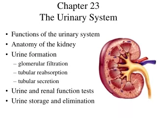
Chapter 23 The Urinary System
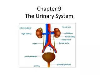
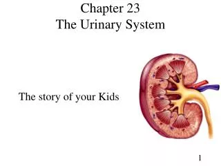
Chapter 23 The Urinary System. The story of your Kids. Urinary system consists of 2 kidneys (kids), each kid has a pipe called the ureter that connects to the bladder, the release hose from the bladder is called the ______ Franklin. Two kidneys. Two ureters. Bladder. Urethra.

Chapter 23 The Urinary System
E N D
Presentation Transcript
Chapter 23The Urinary System The story of your Kids
Urinary system consists of 2 kidneys (kids), each kid has a pipe called the ureter that connects to the bladder, the release hose from the bladder is called the ______ Franklin Two kidneys Two ureters Bladder Urethra
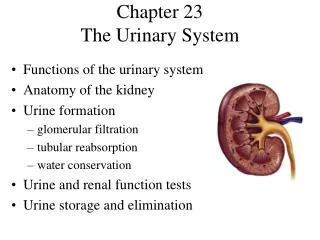
Kidney Location • The kidneys are kidney bean shaped, or… • About the size of your fist • Retroperitoneal-Found posterior to the peritoneum of the abdominal cavity, Right kid lower then left, Why? • Eleventh and twelfth rib partially covers them
The Urinary System • The URINary system’s major job is to produce- URINe • Urine is created from the kids filtering the blood, the result is a sterile waste product called urine • Nephrology (Kidney Study)- is the study of the pathology of the kidneys • Urology (urine study)- study of the male and female urinary system and the male reproductive system
We will concentrate on the kids ability to form urine but they do other things as well: Detoxifies free radicals and drugs Gluconeogenesis- regulates blood sugar pH by regulating H+ and bicarbonate ions Electrolytes regulation in the blood - Na+, K+, Ca++ Erythropoietin secretion Blood pressure regulation- two ways by Renin hormone and Renal resistance Fluid balance- both osmolarity and blood volume by regulating water in the blood Drunk Guys pEE Behind Fences Kidney Functions
Nitrogenous Wastes • Kidneys filter blood plasma, returns useful substances to blood and eliminates waste • Urea • proteinsamino acids NH2 removed forms ammonia NH3, liverconverts to urea • Uric acid • nucleic acid catabolism • Renal failure • azotemia: BUN (Blood Urea Nitrogen), nitrogenous wastes in blood • uremia: toxic effects as wastes accumulate
A sliced kidney reveals two parts: 1.) Renal cortex- outer area 2.) Renal medulla- inner area renal columns, pyramids, papilla Additional anatomy: Renal Capsule- sausage covering Hilus- pushed in center spot where blood vessels, lymphatic vessels, nerves and ureter penetrate the kidney Anatomy of Kidney
Flow- Urine’s Journey • The parenchyma of the kidney contains functional units (about one million) called: • Nephrons- forms urine that passes to- • Collecting ducts- these guys go to the point of the pyramids, then urine flows to:
Of Minors and Majors • Minor calyces to the major calyces- funnel like structures that drain the urine to: • Renal pelvis- large basin that collects the urine and sends it to the ureter to the urinary bladder
Renal artery- main artery entering the renal hilus Renal vein- main vein exiting the kidney Interlobar vessels- pass through the renal columns between the pyramids Arcuate vessels- “ARC” around the distal end of the pyrimids The arcuate vessels send out interlobular vessels that go into the cortex Blood Supply to the Kidneys I
Each nephron receives one afferent arteriole, which divides into a: Glomerulus (little ball)- a ball shaped capillary network Efferent arteriole- reuniting of the glomerulus capillaries that drains the blood out of the glomerulus Peritubular capillaries- further division of efferent arterioles, surround the tubular portions of the nephron Blood Supply to the Kidneys II
The Unique Glomerulus • The glomerulus is unique in two ways: • 1.) Glomerulus capillaries are unique- positioned between two arteries, not (like usual) between an artery and a vein • 2.) The glomerulus is part of the cardiovascular AND urinary systems
Renal CorpuscleGlomerulus and Glomerular capsule (Bowman’s Capsule) Glomerular filtrate collects in capsular space, flows into renal tubule
The Nephron • Functional units of the kidneys • 3 processes: • 1.) Filtering blood • 2.) Returning useful substances • 3.) Removing non-needed substances • NEPHRONS MAINTAINS THE HOMEOSTASIS OF BLOOD AND PRODUCES URINE
Parts of the Nephron • Two portion: • Renal corpuscle • Glomerulus • Glomerular capsule called Bowman’s Capsule • Renal tubule • Proximal convoluted tubule • Loop of Henle (all the good anatomy structures are taken) • Distal convoluted tubule
Renal Tubule • PCT- Proximal convoluted tubule • Nephron loop (Loop of Henle)- U shaped; descending + ascending limbs • Distal convoluted tubule (DCT)
Two Homes for Nephrons • Cortical nephrons (85%) - most of the nephrons • Lie in the cortex mostly • Has SHORT loop of Henle that dips only slightly into the medulla • Juxtamedullary nephrons (15%)- small amount of nephrons • Lie in the cortex BUT close to the medulla • Has LONG loop of Henle that go into the deepest region of the medulla
Parts of the Nephron • Descending limb of the loop of Henle- this part of the loop dips into the medulla • Loop of Henle- connects the proximal to the distal convoluted tubules • Ascending limb of the loop of Henle- this part takes the LOOP (hairpin curve) and returns to the renal cortex • Collecting ducts- the place where the distal convoluted tubules dump • Papillary ducts- converging of many collecting ducts • Don’t Look At Cows Peeing
Glomerular Filtration Rate (GFR) • Filtrate formed per minute • Fluid that enters the capsule space of the glomerulus is called glumerular filtrate. • Filtration fraction = the portion of blood in the afferent arterioles that becomes glumerular filtrate • More then 99% of glumerular filtrate is returned to the blood by tubular reabsorption • About 1-2 quarts is excreted as urine per day
Effects of GFR Abnormalities • GFR, urine output rises dehydration, electrolyte depletion • GFR wastes reabsorbed • GFR controlled by adjusting glomerular blood pressure • autoregulation • sympathetic control • hormonal mechanism: renin and angiotensin
Filtration Membrane • Filtration membrane = A leaky barrier, formed by glomerular capillaries • Permits: filtration of water and small solutes • Prevents: proteins, blood cells from going through
Filtration • Remember, ALL capillaries filter (Starling’s Law) they all have pressure in them • Principle of filtration = Forcing fluids and solutes through a membrane by pressure • But the glomerular capillaries filters more • 3 reasons: • 1.) Large surface area • 2.) Thin and leaky • 3.) High blood pressure- the efferent blood vessels are small, the afferent bigger
The Great Recycling Analogy • Trucks dump into a hopper, where the smaller items passes onto a conveyor belt (glomerular filtration of blood) As the conveyor belt bring the stuff along, workers remove useful items (reabsorption) Other workers place additional larger stuff on the belt (secretion) At the end of the belt all the stuff on the belt goes to the dump (urine)
Antidiuretic Hormone (ADH) • ADH- a hormone that stops water loss • ADH Is inhabited by alcohol the result is dilute (clear) but plentiful urine • The rate at which H2O is lost from the body is dependent upon ADH • When ADH is high, urine is concentrated • When ADH is low, dilute urine is produced
Kidney Tests • Blood tests • BUN- blood urea nitrogen, the result of protein metabolism, increased BUN is bad • Indications are renal disease or obstruction of the urinary tract • Urinalysis (UA)- evaluation of the chemical of the urine • Microscopic and DIP-STICK is used to evaluate urine
Dip Stick I • (Look to your right) • A thin piece of plastic with small boxes of chemical that is placed in urine and tells: • pH- normal avg. 6 • Specific gravity- urine weight compared to water, should be 1.001-1.035, just slightly heavier then water, but not much more or the urine will have substances in it
Dip Stick II • Albumin- a protein in the blood, big molecule, if found in large amounts then the membranes of the kidney are damaged to let it through • Glucose- If too much glucose is in the blood (usually above 300 ppm) it will spill into the urine, no glucose should be found in the urine, suspect diabetes • Blood- if found in urine it may be due to menstrual blood, trauma to the genitals or kidney stones • Leukocytes- possible infection or not a “clean catch”, if infection is suspected must be correlated with other tests
Dip Stick III • Ketones- Byproduct from fatty acid metabolism, fasting, anorexia and high fat/ low carbohydrates diets can raise ketones. Also seen in diabetes, correlate with ________ * • Bilirubin- excessive of RBCs being destroyed by macrophages • Urobilogen- Anemia, liver problems, mono
Micro/Macroscopic Urine • MACRO- Urine’s color should be yellow to amber, (red urine? Usually not blood but _____ • Turbidity- Urine should be transparent after voiding • Odor- Should not be sweet smelling • If it is sweet smelling then suspect diabetes • MICRO- Casts are tiny tubes found in the urine • Microbes- E. Coli, Candida
Urine’s Journey Out… • Urine drains from the calyces (minor then major) to the renal pelvis into the ureters and then into the urinary bladder and out it goes through the urethra • Ureters- peristaltic contraction in the walls of the ureters push urine to the bladder • A foot long • At the end of the ureters is a hole that acts like a valve that shuts when the bladder gets full, preventing back flush • mucosa - transitional epithelium • lumen very narrow, easily obstructed
…The Journey Continues • Urinary bladder- hollow, muscular organ just posterior to the symphysis pubis. • The bladder is like a triangle pointing down, the top superior and lateral points are where the two ureters enter the bladder, mucosa: transitional epithelium • The Urethra drains the bladder at the inferior point of the triangle, rugae: relaxed bladder wrinkled • The urethra has circular muscle fibers that form the internal urethral sphincter • The urethra has another sphincter below the internal sphincter called the external sphincter, it is composed of skeletal (Hold it!) voluntary muscle • Capacity: moderately full = 2 cups, maximum = 3 ½ cups
Female Urethra • Short, purpose- void urine • External urethral orifice • between vaginal orifice and clitoris • Internal urethral sphincter • smooth muscle, involuntary control • External urethral sphincter • skeletal muscle, voluntary control
Male Bladder and Urethra • Long, duo purpose = void urine & Ejaculate • Internal urethral sphincter • External urethral sphincter • 3 regions • prostatic urethra • during orgasm receives semen • membranous urethra • passes through pelvic cavity • spongy urethra in penis
Voiding Urine – Micturition • Micturition- urination, voiding, number one, peeing • When the bladder gets full, stretch receptors initiate the spinal nerves to open the sphincters • Because the external sphincter is skeletal muscle we can over ride this reflex and “Hold it!” • Valsalva maneuver • aids in expulsion of urine by pressure on bladder • can also activate micturition reflex voluntarily • A lack of the ability to hold it is called incontinence, failure to void completely is called retention
Waste Management • The urinary system is concerned with getting rid of waste • Other organs join in with this task: • Blood/ lymphatic system • Liver • Gall bladder- holds waste accumulated from liver • Skin • GI tract
URINARY DIS-EASE I • UTI- Urinary tact infection- more common in females due to short urethra • Bright’s disease- inflammation of glomeruli of kidney, strep infection also called glomerulonephritis • Renal disease- chronic and acute, decreased glomerular filtration rate • Kidney stones- majority are calcium stones, pain more from urinary backup than stone pressing into ureter
URINARY DIS-EASE II • Urethritis- inflamed urethra from UTI usually • Cystitis- inflammation of bladder from UTI or honeymoon • Pyleonephritis- inflammation of kidneys usually from bladder infection going up commonly called Kidney infection • Polycystic kidney disease- cysts of the kidneys, inherited, causes renal failure shortly after birth
URINARY DIS-EASE III • Enuresis- (en-yoo-RE-sis)- bed wetting • Pyelogram (PI-e-lo-gram)- X-ray of the kids after iodine injection • Polyuria- peeing too much, a bird’s urine