Download

1 / 14
140 likes | 161 Views
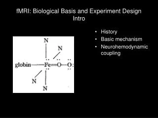
fMRI: Biological Basis and Experiment Design Intro. History Basic mechanism Neurohemodynamic coupling. NMR - MRI - fMRI timeline. 1922 Stern-Gerlach Electron spin. 1952 Nobel prize Felix Bloch, Edward Purcell NMR in solids. 1993 Seiji Ogawa, et al. BOLD effect. 1902 Pieter Zeeman

E N D
fMRI: Biological Basis and Experiment DesignIntro • History • Basic mechanism • Neurohemodynamic coupling
NMR - MRI - fMRI timeline 1922 Stern-Gerlach Electron spin 1952 Nobel prize Felix Bloch, Edward Purcell NMR in solids 1993 Seiji Ogawa, et al. BOLD effect 1902 Pieter Zeeman Radiation in a magnetic field 1937 Isidor Rabi Nuclear magnetic resonance 1973 Paul Lauterbur, Peter Mansfield NMR imaging 1936 Linus Pauling Deoxyhemoglobin electronic structure
... ... PNAS22(4):210-216
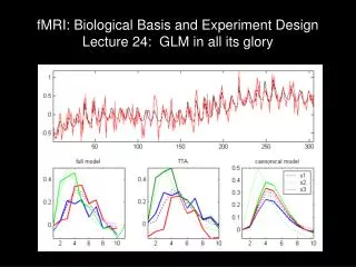
Basic BOLD “... blood oxygenation level-dependent (BOLD) contrast: a change in the signal strength of brain water protons produced by the paramagnetic effects of venous blood deoxyhemoglobin.” –Ogawa et al. 1993 Signal inversely proportional to deoxyhemoglobin concentration • CBF = cerebral blood flow • increased CBF increases signal strength • CBV = cerebral blood volume • increased venous blood volume decreases signal strength • CMRO2 = cerebral metabolic rate of oxygen • increased CMRO2 decreases signal strength
Neural layers and vasculature Duvernoy, Delon & Vanson, Brain Res. Bull., 1981
What is neural activity? Neural activity: - increased oxygen consumption (CMRO2) - increased need for glucose (CMRglc)
5m What does blood flow have to do with neural activity? Upstream arteries: - increase flow (CBF) brings oxygen and glucose Downstream veins: - increased blood volume (CBV) - decreased deoxyhemoblogin concentration Neural activity: - increased oxygen consumption (CMRO2) - increased need for glucose (CMRglc)