Download
1 / 35
370 likes | 527 Views
Explore the functions of veins, central venous pressure, venous return determinants, and more. Educational resources provided for comprehensive learning.

E N D
Intended learning outcomes (ILOs) After reviewing the PowerPoint presentation and the associated learning resources, the student should be able to: Discuss functions of the veins as blood reservoirs. Describe measurement of central venous pressure (CVP) and state its physiological and clinical significance. State determinants of venous return and explain how they influence venous return. Define mean systemic filling pressure, give its normal value and describe the factors which affect it. Explain the effect of gravity on venous pressure and explain pathophysiology of varicose veins. Describe vascular and cardiac function curves under physiological and pathophysiological conditions.
Learning Resources Guyton and Hall, Textbook of Medical Physiology; 13thEdition; Unit IV-Chapter 20. Linda Costanzo, Physiology, 5th Edition; Chapter 4. Ganong’sReview of Medical Physiology; 25th Edition; Section V; Chapters 30 and 31.
Major components of the cardiovascular system HEART • Veins are thin walled vessels with relatively large lumen. • They can accommodate large changes in blood volume with little change in pressure until a limit is reached. • Beyond the limit, any change in volume results in change in pressure. VEINS CAPACITY VESSELS ARTERIES (LOW COMPLIANCE) CAPILLARIES
Veins • Structure: • all 3 layers are present, but thinner than in arteries of corresponding size (external diameter). • Veins have paired semilunar, bicuspid valves to restrict backflow in lower extremities: • In varicose veins, blood pools because valves fail causing venous walls to expand.
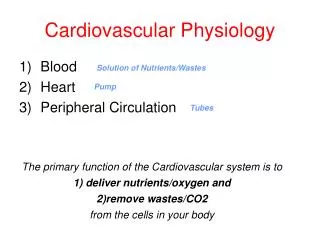
Veins are the capacitance vessels of the circulation. • Veins function as variable reservoir of blood with ≈2/3 blood volume contained in them. • They collect and return blood to the heart. Thus, they are important in venous return to the heart. Functions of veins
Veins serve as blood reservoirs Normally all the blood is circulating all the time. When the body is at rest and many of the capillaries are closed, the capacity of the venous reservoir is increased as extra blood bypasses the capillaries and enters the veins. When this extra volume of blood stretches the veins, the blood moves forward through the veins more slowly because the total cross sectional area of the veins has increased as a result of the stretching. Therefore, blood spends more time in the veins. As a result of this slower transit time through the veins, the veins are essentially storing the extra volume of blood because it is not moving forward as quickly to the heart to be pumped out again. When the stored blood is needed, such as during exercise, extrinsic factors reduce the capacity of the venous reservoir and drive the extra blood from the veins to the heart so that it can be pumped to the tissues.
Venous return (VR) • Venous return (VR) is the flow of blood back to the heart. • Under steady-state conditions, venous return must equal cardiac output (CO) when averaged over time because the cardiovascular system is essentially a closed loop. Otherwise, blood would accumulate in either the systemic or pulmonary circulations. Venous return is determined by the difference in pressure between the venous pressure nearest to the tissues (mean systemic filling pressure; mean circulatory pressure; MCP) and the venous pressure nearest to the heart (CVP).
CVP: is the venous pressure in the right atrium and the big veins of the thorax (= right atrial pressure (RAP) = jugular venous pressure). Venous pressure is measured with a catheter inserted in the central venous system, usually SVC. The normal range of the CVP = 0 - 4 mm Hg. It is the force responsible for cardiac filling. CVP is used clinically to assess hypovolaemia and during IV transfusion to avoid volume overloading. CVP is raised in right-sided failure. Central venous pressure (CVP)
Mean systemic filling pressure Mean circulatory pressure; MCP • It is the pressure nearest to the tissues. • Its normal value is about 7 mm Hg. • The value for right atrial pressure at which venous return is zero is called the mean systemic filling pressure. It is the point at which the vascular function curve intersects the X-axis (i.e., where venous return is zero and right atrial pressure is at its highest value). • It is affected by: • Blood volume (it is directly proportional to blood volume). • Venous capacity (it is inversely proportional to the venous capacity).
Mean circulatory pressure; MCP MCP is determined by blood volume and venous capacity Unstressed Volume Stressed Volume • The unstressed volume is the volume of blood in the vasculature that produces no pressure. • The stressed volume is the volume that produces pressure by stretching the elastic fibers in the blood vessel walls. Infusion VOLUME MCP Normal 7- MCP (mmHg) VOLUME MCP Hemorrhage 1 2 3 4 5 6 BLOOD VOLUME (L)
Mean circulatory pressure; MCP VENOCONSTRICTION Unstressed Volume Stressed Volume Normal 7- MCP (mmHg) 1 2 3 4 5 6 BLOOD VOLUME (L)
Mean circulatory pressure; MCP VENODILATION Unstressed Volume Stressed Volume Normal 7- MCP (mmHg) 1 2 3 4 5 6 BLOOD VOLUME (L)
Determinants of venous return Blood volume Venous capacity Sympathetic activity Skeletal muscle activity Venous valves Respiratory activity Gravity.
Determinants of venous return • 1. Blood volume: • At constant venous capacity, as the blood volume → the MCP → VR. • At constant venous capacity, as the blood volume↓→ the MCP ↓→ ↓VR.
Determinants of venous return • 2. Venous capacity: is the volume of the blood that the veins can accommodate. • It depends on the distensibility of the vein walls and the influence of any externally applied pressure squeezing inward on the veins. • At a constant blood volume, as the venous capacity → more blood spends a longer time in the veins instead of being returned to the heart → ↓ the effective circulating volume → ↓ VR. • At a constant blood volume, as the venous capacity → the MCP ↓ → ↓ VR. • As the venous capacity ↓ → VR.
Determinants of venous return • 3. Sympathetic activity: • Venous smooth muscle is profusely supplied with sympathetic nerve fibers. • Sympathetic stimulation → venous vasoconstriction → modest in mean systemic filling pressure (MCP) → VR. • Sympathetic stimulation → ↓venous capacity→ VR. • What is the effect of venoconstriction on the resistance to flow? • The veins normally have such a large diameter that the moderate vasoconstriction accompanying sympathetic stimulation has little effect on resistance to flow.
Determinants of venous return • 4. Skeletal muscle activity: • Skeletal muscle contraction → external venous compression → ↓venous capacity→ VR (This is known as skeletal muscle pump). • Skeletal muscle activity also counter the effects of gravity on the venous system.
Determinants of venous return • 5. Venous valves: • These valves permit blood to move forward towards the heart but prevent it from moving back toward the tissues. • These valves also play a role in counteracting the gravitational effects of the upright posture.
Skeletal muscle pump is ineffective when the venous valves are incompetent. • Chronically raised pressure in the veins leads to pathological distension of the veins (varicose veins). • Increased capillary filtration leads to swelling (edema) with trophic skin changes and ulceration (venous ulcers). Venous incompetence
6. Respiratory activity (respiratory pump; thoracic pump): • As the venous system returns blood to the heart from the lower regions of the body, it travels through the chest cavity. The pressure in the chest cavity is 5 mm Hg less than atmospheric pressure. • The venous system in the limbs and abdomen is subjected to normal atmospheric pressure. • Thus, an externally applied pressure gradient exists between the lower veins and the chest veins, promoting venous return (this is known as the respiratory pump). • What is the effect of Valsalva maneuver on venous return? Determinants of venous return
Determinants of venous return What is the effect of Valsalva maneuver on venous return?
Determinants of venous return 7. Effect of gravity on venous return • Venous compliance is high and the veins readily expand with blood. Thus, upon standing from the supine position, most of the blood volume shift occurs in the veins. • Therefore, venous volume and pressure become very high in the feet and lower limbs when standing. This shift in blood volume decreases thoracic venous blood volume and therefore CVP decreases. • This decreases right ventricular filling pressure (preload), leading to a decline in stroke volume by the Starling mechanism. • Left ventricular stroke volume also falls because of reduced pulmonary venous return (decreased left ventricular preload). This causes CO and mean arterial pressure (MAP) to fall. • If arterial pressure falls appreciably upon standing, this is termed orthostatic or postural hypotension. • This fall in arterial pressure can reduce cerebral blood flow to the point where a person might experience syncope (fainting).
There is an inverse relationship between venous return and right atrial pressure (RAP). • The inverse relationship is explained as follows: • Venous return back to the heart, like all blood flow, is driven by a pressure gradient. The lower the pressure in the right atrium, the higher the pressure gradient the greater the venous return. • Thus as RAP increases, this pressure gradient decreases and venous return also decreases. • The knee (flat portion) of the vascular function curve occurs at negative values of RAP. At such negative values, the veins collapse. This collapse impedes blood flow back to the heart. Thus, although the pressure gradient has increased (i.e., as RAP becomes negative), venous return levels off because the veins have collapsed. Venous return curve Vascular function curve MCP RAP
Vascular function curve 10- 5- 0- Blood Volume or Venoconstriction VENOUS RETURN (L/min) MCP MCP Blood Volume or Venodilation -4 0 +4 +8 RAP (mmHg) • If blood volume increases, the amount of blood in the unstressed volume will be unaffected, but the amount of blood in the stressed volume will increase. When stressed volume increases, mean systemic pressure increases and the vascular function curve and its intersection point with the X-axis shift to the right. The same effect is seen with venoconstriction. • If blood volume decreases, then stressed volume decreases, mean systemic pressure decreases, and the vascular function curve and its intersection point with the X-axis shift to the left. The same effect is seen with venodilation.
Vascular function curve Vasodilation 10- 5- 0- VENOUS RETURN (L/min) TPR TPR Vasoconstriction -4 0 +4 +8 RAP (mmHg) • When the TPR is decreased, for a given RAP, venous return is increased. In other words, decreased resistance of the arterioles (decreased TPR) makes it easier for blood to flow from the arterial to the venous side of the circulation and back to the heart. • When the TPR is increased, for a given RAP, venous return is decreased. In other words, increased resistance of the arterioles (increased TPR) makes it more difficult for blood to flow from the arterial to the venous side of the circulation and back to the heart.
Combining cardiac and vascular function curves • When cardiac output and venous return are plotted simultaneously as a function of RAP, they intersect at a single value of RAP. • At this one value of RAP, cardiac output equals venous return and, by definition, is the steady state operating point of the system. • That one value of RAP satisfies both cardiac output and venous return relationships. 15- CARDIAC OUTPUT or PERIPHERAL BLOOD FLOW [Venous Return] (L/min) 10- 5- -4 0 +4 +8 RAP mmHg
Combining cardiac and vascular function curves
Combining cardiac and vascular function curves 1. Effects of changes in blood volume • Increases in blood volume as a result of transfusion of a large fluid volume into the circulationincrease the amount of blood in the stressed volume and, therefore, increase the mean systemic pressure. • This results in shifting of the vascular function curve to the right. • The cardiac function is not altered with changes in blood volume. • In the new steady state, the cardiac and vascular function curves intersect at a new point at which venous return and the cardiac output are increased. The RAP is increased.
Combining cardiac and vascular function curves 2. Inotropic effects • Positive inotropic agents produce an increase in contractility, an increase in stroke volume, and an increase in cardiac output for any level of RAP. Thus, the cardiac function curve shifts upward (counter-clockwise), but the vascular function curve is unaffected. • Thus, there will be substantial increases in the cardiac output and venous return , while the RAP is decreased. • The opposite is true with negative inotropic agents.
Combining cardiac and vascular function curves 3. Increased sympathetic nervous system tone • Increased SNS tone rotates the cardiac function curve counter-clockwise due to its boosting of the cardiac contractility and heart rate. • Additionally, SNS stimulation reduces venous vascular compliance and thus causes venoconstriction which increases the "Mean Systemic Pressure", shifting the vascular function curve to the right. • The combined effect of these changes is a substantial increase in the cardiac output and venous return without a large change in the RAP.
Combining cardiac and vascular function curves 4. Effects of changes in TPR • Changes in TPR alter both curves. The cardiac function curve changes because of a change in afterload (arterial blood pressure), and the vascular function curve changes because of a change in venous return. • With increased TPR, there is a substantial decrease in the cardiac output and venous return with almost no change in the RAP. The opposite is true when the TPR is decreased.
Combining cardiac and vascular function curves in heart failure
Intended learning outcomes (ILOs) After reviewing the PowerPoint presentation and the associated learning resources, the student should be able to: Discuss functions of the veins as blood reservoirs. Describe measurement of central venous pressure (CVP) and state its physiological and clinical significance. State determinants of venous return and explain how they influence venous return. Define mean systemic filling pressure, give its normal value and describe the factors which affect it. Explain the effect of gravity on venous pressure and explain pathophysiology of varicose veins. Describe vascular and cardiac function curves under physiological and pathophysiological conditions.