Download
1 / 51
510 likes | 652 Views
This guide covers signalment, conditions, and examinations related to cardiac and pulmonary issues in small animals. Learn to differentiate between syncope and seizures, understand causes of cough, and master stethoscope use for auscultation.

E N D
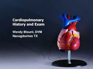
Cardiopulmonary History and Exam Wendy Blount, DVM Nacogdoches TX
Signalment Age • Congenital disease • young • Myxomatous valvular Disease • old • Exceptions • Cavalier King Charles Spaniels • PDA • Reverse PDA
Signalment Breed • Boston Terrier • Cavalier • Cocker Spaniel • Boxer • Doberman • English Bulldog • Golden Retriever HBT, CollapsingTr CVD DCM, PS, PDA, 3rdAV HBT, PS, SAS DCM, Boxer CM, ASD DCM (Arrhythmia?) SAS, PS, CVD SAS
Signalment Breed • Great Dane • GSD • Irish Setter • Irish Wolfhound • Keeshond • Labrador • Maine Coon • Newfoundland DCM, CVD PRAA, SAS, PDA PRAA DCM TOF (define), MVD TVD HCM DCM, SAS
Signalment Breed • Persian/Himalayan • Pointer • Poodle • St Bernard • Samoyed • Schnauzer • Springer Spaniel • Yorkie HCM PRAA, SAS CVD, PDA, CB DCM ASD, PS SSS, CVD, PS, CB VSD CVD, CB, CT
History - Collapse How can you tell the difference between seizure and syncope? • Urination/defecation/vocalization/paddling • Stiff/opisthotonus or flaccid • narcolepsy • Twitching and muscle fasciculations • Cyanosis, pallor • Abnormal behavior before and after • Duration of stiffness/opisthotonus Many times, you can’t (especially when short)
History - Collapse What causes syncope? • Bradyarrhythmia • 3rd degree heart block (define) • Sick sinus syndrome (define) • Period of asystole • Sick sinus syndrome • Vagal surge (examples) • Abdominal dz & retching • Intubation (brachycephalic)
History - Collapse What causes syncope? • Tachyarrhythmia burst • Vtach (causes) • BCM • Myocarditis • Myocardial hypoxia • Abdominal pathology (spleen) • Supraventricular tachycardia (SVT) (define) • Re-entry pathway (define) • Atrial fibrillation (Afib) • SSS (3 ways)
History - Collapse What causes syncope? • Obstruction of a great vessel or heart chamber • Thrombus • neoplasia • Increased oxygen demand can not be met due to severe cardiovascular or pulmonary disease • AKA Exercise intolerance
History - Cough How can you tell the difference between cardiac and respiratory cough/dyspnea? • Honking cough • Soft moist cough • Dry hacking cough • Coughing/gagging up white foamy fluid • Coughing up blood tinged fluid • Cough when drinking water • Exercise induced cough • Presence of a murmur (big dog, little dog) Many times, you can’t without PE/diagnostics
History - Cough Cough on tracheal palpation • Any dog or cat will cough a few times on vigorous tracheal palpation • Prolonged coughing after tracheal palpation often indicates pathology (cardio or resp?) • Prolonged coughing equally likely with airway disease and cardiovascular disease
History - Cough Dogs vs Cats • Coughing cats • much more likely to have respiratory disease than heart failure • Cats with heart failure more often present with acute and severe dyspnea • Some owners can find it difficult to distinguish vomiting from coughing • Coughing dogs can have either or both
Exam – Stethoscopes • Ear pieces fit snugly in the ears • Angle fits your ear canals • Poor fit, and you’ll miss low intensity murmurs • Tubing longer than 18 inches will dampen sounds Electronic stethoscopes (microphone based) • Difficult to distinguish heart from lung sounds • Difficult to distinguish patient from background noise • Meditron sensor based scope eliminates problems • Connect to computer & record for PCG consult
Exam – Stethoscopes Pediatric stethoscope • For cats and small dogs • Will distort and decrease sound intensity if used on a medium or large dog Adult stethoscope • For medium to large dogs • Won’t localize murmurs properly in cats and small dogs
Exam – Stethoscopes Diaphragm • Filters out low frequency sounds to hear high frequency sounds better • Press firmly against the chest Bell • For low frequency sounds (S3 S4 in dogs) • Press gently against the chest
Auscultation Minimizing patient noise • Panting, whining – close mouth, occlude nostrils • Purring • Aversives – turn water on, show another animal • Gentle pressure on the larynx • Cotton ball with alcohol to the nose • Sometimes sedation is needed (chart) • Acepromazine 0.0125-0.025 mg/lb, maximum 1 mg per dog • Butorphanol 0.1 mg/lb or buprenorphine 0.01-0.02 mg/kg • IV the fastest and most profound
Auscultation • Patient is standing in a quiet place • Lateral recumbency and listen from bottom if muffled • Firm pressure with the diaphragm to avoid hair noises • get comfortable ausculting heart and palpating pulses at the same time • Listen for at least 2 minutes • Heart - R and L apex, L bases • L armpit • Lungs – RDCr, RDCd, RVentral, LDCrd, LDCd, LVentral
Auscultation • Is the murmur hemodynamically significant? • Prolonged and loud - yes • pansystolic - yes • Diastolic - yes • Low intensity, early systolic – maybe not so much Loudness is not necessarily correlated to presence of heart failure
Auscultation – Lung Sounds • Snaps crackles and wheezes (cardio or resp?) • More likely respiratory in dogs (audio) • Not very sensitive for pulmonary edema • Beware similar hair rubbing noises • Pleural/pericardial Rubs (audio) • Dull/absent lung sounds (dog vs cat) (causes) • Lung consolidation • Pneumothorax • pleural effusion • Harsh lung sounds with no murmur in cat • think asthma or heartworm disease
Auscultation - Heart Sounds Normal Heart Sounds
Auscultation - Heart Sounds Normal Heart Sounds • HS1 • AV Valves close • Beginning of systole • HS2 • Semilunar valves close • end of diastole • Tachycardia – which is which? • S2 shorter and higher frequency
Auscultation - Heart Sounds • Variable intensity HS1 • arrhythmia • Louder HS1 (AV slamming) • Young, narrow chested dogs • Increased sympathetic tone • Anemia • Fever • Hypertension • Advanced mitral valve disease
Auscultation - Heart Sounds • Quieter HS1 (AV softly closing or muffled) • Obesity, barrel chested dogs • Myocardial failure • Pronounced 1st degree heart block • hypervolemia
Auscultation - Heart Sounds • Louder HS2 (SL slamming) • Hyperthyroidism • Fever, anemia • Heartworm Disease • Cor pulmonale (define) • Quieter HS2 (SL softly closing) • Myocardial failure (DCM, severe MR)
Auscultation - Heart Sounds Third Heart Sound (Gallop) HS3 (1-2-3) HS4 (4-1-2) Split S2 Systolic Click Summation Gallop (4-1-2-3)
Auscultation - Heart Sounds Third Heart Sound HS3 – protodiastolic gallop (1-2-3) • Rapid LV filling – early diastole (audio) • PMI R or L apex – low frequency • At maximal mitral opening (E point on echo) • stiff LV or large diastolic volume • HCM, RCM, DCM, severe MR • Indicates myocardial failure • Usually a bad mamma jamma
Auscultation - Heart Sounds Third Heart Sound HS4 – presystolic gallop (4-1-2) • Atrial contraction - Late diastole (audio) • PMI R or L apex, low frequency • Stiff LV (HCM) • Increased afterload • 3rd degree AV block • Myocardial failure (DCM, bad MR) • Sometimes heard in normal cats & giant dogs • not necessarily a bad mamma jamma
Auscultation - Heart Sounds Third Heart Sound • Split 2nd Heart Sound • PMI right heart base (left side) • AoV PV don’t close at same time (PV later) • Reverse PDA • Pulmonary hypertension (HWDz) • Severe RBBB • relative PS of right to left shunts (ASD) • normal variation in large dogs (audio)
Auscultation - Heart Sounds Third Heart Sound Systolic Click • Very sharp, high frequency click of Mitral valve prolapse, in early CVD • Snapping of the chordae tendinae as they go taught • PMI left apex • Mid-Systolic (audio) • May me accompanied by a systolic murmur • Early, late, or holosystolic • Often years until CHF develops
Auscultation – 3 Heart Sounds How Can you tell the difference? Does in Matter? • Systolic less likely pathogenic • Systolic Click sounds sharper • Diastolic more likely pathogenic How Can you tell if systolic/diastolic? • Pulses happen during systole How Can you tell if HS3 or HS4? • Can’t tell if heart rate is > 160-180 • just do a cardio work-up
Auscultation – Heart Sounds PMI 1 – left apex (MV) 2 – left base (AoV) 3 – right base (PV) 5 4 – right apex (TV) 5 – left armpit (PDA)
Auscultation – Heart Sounds PMI (Point of Maximal Intensity) Left Apex – at palpable apical beat (HS loudest?) • S1 - MR (audio) Left Base – cranial & dorsal (HS?) • S2 - SAS (audio) • S2 - Ao endocarditis (audio) Right Base (left side) (HS?) • S2 - PS (audio) Left Axilla (HS?) • continuous - PDA (audio) Right Apex (HS?) • S1 - TR
Auscultation – Heart Sounds Muffled Heart Sounds (causes) • Pleural, Pericardial effusion (*difference*) • Diaphragmatic hernia, thoracic masses • obesity What besides cardiac disease can cause a pathologic murmur? • Anemia, hypoproteinemia Why do puppies have innocent murmurs? • Musical • Larger SV relative to great vessel size • Lower PCV and plasma proteins • Artifact – high frequency breath sounds
Auscultation – Murmur Grade Grade 1 • Heard in a very quiet room, concentrating Grade 2 • easily heard on the PMI Grade 3 • Moderately loud Grade 4 • Very loud over much of the chest Grade 5 • Heard with edge of stethoscope on chest, palpable thrill Grade 6 • Heard with stethoscope off chest, palpable thrill
Auscultation – Murmur Grade High grade murmurs are more likely to be associated with severe disease Severe disease can also be present with low grade murmur • Occasionally no auscultable murmur in the cat • DCM • ASD • VSD • Reverse PDA (right to left shunting)
Auscultation – Murmurs holosystolic • Starts at the end of S1 • Ends at the start of S2 • Murmur between the heart sounds
Auscultation – Murmurs pansystolic • Starts at the beginning of S1 • Ends at the end of S2 • Just hear the murmur with no distinct HS
Respiratory Sinus Arrhythmia • Heart rate increases during respiration • Due to increased vagal tone • Normal variation in dogs (not cats) • If present, heart failure is not likely • Increased sympathetic tone overrides • Pronounced in disease processes of increased vagal tone • Increased CSF pressure • Chronic respiratory disease • Thoracic or abdominal disease
Respiratory Sinus Arrhythmia • DDx • Afib with a normal ventricular rate • Frequent APCs or VCPs • Intermittent SSS None of these vary consistently with the respiratory cycle • RSA is regularly irregular • Others are usually irregularly irregular
Physical Exam – Ascites • most common cause of cardiogenic ascites in cats (?) • TVD • Tap and do fluid analysis to distinguish between transudate, modified transudate and exudate (handout) • Usually accumulates slowly, though owners often don’t notice until huge • If truly does develop over days, think pericardial tamponade
Exam – Mucous Membranes Cyanosis • > 4 g/dL of deoxygenated Hb in the blood • Severely anemic animals don’t turn blue • Even with life threatening hypoxia • Differential cyanosis (define) • Front of body pink, back of body blue • Reverse PDA, FATE (why rPDA)(how to diagnose?) • Compare pulse oximetry or blood gases from front of body with rear of body • Weak or no femoral pulses, pain, paresis with FATE
Exam – Pulses Technique • Occlude the pulse • Then slowly release pressure until maximum pulse is detected Pulse Pressure = Systolic – Diastolic • Femoral pulse usually not palpable when MAP <50mmHg • Dorsal pedal pulse not palpable when SAP <80mmHg
Exam – Pulses Bounding Pulses (water hammer) • Increased systolic pressure (increased SV) (causes) • Aortic regurgitation • Severe bradycardia • Thyrotoxicosis (define EF, FS) • Fever • Anemia/hypoproteinemia • decreased diastolic pressure (diastolic runoff) • PDA • AV fistula • Aortic regurgitation (most common cause) • Aortic endocarditis > SAS
Exam – Pulses Weak Pulses • Severely decreased SV – severe HF • Acutely decreased SV – hypovolemia • Decreased peripheral vascular resistance (shock) • Decreased arterial compliance (hypertension) Pulse peaks slowly and late in systole • Pulsus parvus et tardus (cause) • Severe SAS
Exam – Pulses Short, Brisk Pulses (snappy) • Short, fast systole • Compensated MR (what happens to FS with MR) Pulse weak or absent during inspiration • Pulsus paradoxus • Systolic pressure falls during inspiration • With pronounced respiratory sinus arrhythmia • Exaggerated by pericardial effusion
Exam – Pulses Alternating Weak and Normal Pulses • Pulsus alternans • Severe myocardial failure (define MF vs CHF) (causes) • DCM • RCM (define) • End stage valvular disease • Prolonged tachyarrhythmia or tachycardia
Exam – Pulses Pulse Deficits (heart beat generates no pulse) • VPCs • Atrial fibrillation with VPCs • Tachyarrhythmia (inadequate filling) • Every other heart beat has a pulse deficit • Pulsus bigeminis • Caused by ventricular bigeminy (define) Totally chaotic heart sounds and pulses (audio) • Lots of multiform VPCs • Atrial fibrillation
Exam – Jugular Veins • Clip or wet the fur over the jugular veins • Evaluate sitting or standing (not sternal) • Jugular Distension(causes) • suggests increased RA pressure (normal dogs cats?) • 2-3 cm H20 in cats, 5-8 cm H20 in dogs • Or less often jugular or caval occlusion • Jugular Pulse(normal dogs cats) • 5-8cm dorsal to RA in dogs, 2-3 cm in cats • Too high indicates increased right heart pressure • If abnormalities above not noted, occlude at thoracic inlet, and release • Hepatojugular reflux
Exam – Jugular Veins Jugular distension, high pulse, +HJR (causes) • Jugular/caval occlusion • Heartworm disease • External mass (cyst, abscess, granuloma, neoplasia) • Thrombus(causes) • Decreased RV compliance • RV hypertrophy • PS, TOF, pulmonary hypertension • Restrictive CM • RVOT obstruction • Heartworm disease, neoplasia, thrombus
Exam – Jugular Veins Jugular distension, high pulse, +HJR • RV volume overload • TR with RHF • VSD • HWDz • Compression on the RV, so it can’t fill • Pericardial effusion • constrictive pericarditis • Pericardial mass Evaluation of hepatic & splenic veins on US are even more sensitive for increased RV pressure