Download
1 / 51
720 likes | 1.97k Views
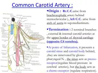
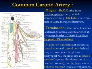
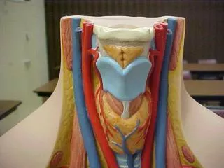
Right external carotid artery. Right internal jugular vein. Right superior carotid artery. Right common carotid artery. Thyroid cartilage. Cricoid cartilage. Thyroid gland. Sclaneus anterior muscle. trachea. Left internal carotid artery. Sternocleidomastoid muscle. Hyoid bone.

E N D
Right external carotid artery Right internal jugular vein Right superior carotid artery Right common carotid artery Thyroid cartilage Cricoid cartilage Thyroid gland Sclaneus anterior muscle trachea
Left internal carotid artery Sternocleidomastoid muscle Hyoid bone Preepiglottic fat pad Left common carotid artery Left internal jugular vein Cricothyroid muscle Left superior thyroid artery Inferior belly of omohoid muscle
Left subclavian artery Right subclavian vein clavicle Right brachiocephalic vein Left subclavian vein Left superior pulmonary vein Superior vena cava aorta Left pulmonary artery Right lung Left primary bronchus Tracheal bifurcation esophagus
6th rib Bronchus of left inferior lobe 7th rib External oblique muscle diaphragm
Abdominal part of esophagus Celiac trunk Right adrenal gland Inferior vena cava ureter Iliac crest
Diaphragm (costal part) Diaphragm (lumbar part) Renal pelvis Renal vein Right testicular artery Right common iliac vein
Parietal pleura Left adrenal gland spleen Splenic hilum Abdominal aorta Left kidney Left ureter Left testicular vein Quadratus lumborum muscle Left testicular artery
Transversus abdominus muscle Psoas major muscle Psoas major muscle Iliacus muscle Internal oblique muscle External oblique muscle Rectus abdominal muscles
Right common iliac artery Median sacral artery Sigmoid colon rectum Urinary bladder
Urinary bladder Femoral artery Lymph vessel Great saphenous vein
glabella Frontal region Temporal region Orbital region Superior palpebra Zygomatic region Infraorbital region Mentolabial sulcus
Superior frontal sulcus Cerebral artery Medial frontal gyrus Sphenoid bone Frontal bone Supraorbital foramen Ocular bulb Lacrimal sac External carotid artery tongue Sublingual gland Genioglossus muscle
Left telecenphalic hemisphere Parietal bone Temporal bone Sphenoid bone Muscle of uvula Nasal bone Posterior belly of digastric muscle Sublingual gland tongue External jugular vein mandible First incisor
Temporalis muscle Lacrimal gland Zygomatic bone Facial artery Styloglossus muscle External carotid artery Submandibular gland Stylohyoid muscle Mylohyoid muscle
External acoustic meatus Triangular fossa tragus antihelix helix antitragus lobule
Falx of cerebellum Occipital pole Tentorium of cerebellum cerebrum Cerebellar tonsil cerebellum Occipital bone Medulla oblongata Transverse process of atlas C1, Atlas Cervical spinal ganglion Cervical spinal ganglion
Cervical spinal ganglion Sclaneus posterior muscle Posterior root of spinal nerve Posterior median sulcus Transverse process of 6th cervical vertebra Right vertebral artery Transverse process of 7th cervical vertebrae T1
1st thoracic vertebra Posterior inter- transverse muscle 2nd thoracic vertebra Thoracic spinal ganglia Spinal cord dura mater Parietal pleura Posterior internal vertebral venous plexus
Spinal arachnoid Thoracic ganglia Intercostal artery 6th rib Intercostal vein Spinal cord (thoracic part) Intercostal nerve Thoracic vertebrae
Facet of superior articular process of thoracic vertebrae Spinous process of 7th thoracic vertebrae Vertebral canal Spinal cord (grey matter) Intervertebral disc Spinal cord (white matter) Dura mater of spinal cord Epidural cavity 10th thoracic nerve
12th rib Fila radicularia Medullary cone Transverse process of 1st lumbar vertebrae Quadratus Lumborum muscle 2nd lumbar vertebrae Psoas major muscle Lateral lumbar intertransverse muscles Lateral lumbar intertransverse muscles Terminal filament
12th rib Transverse process of 1st lumbar vertebrae Psoas major muscle Lateral lumbar intertransverse muscles Terminal filament 4th lumbar vertebrae Posterior roots of lumbar and sacral nerves
Lumbar spinal ganglion 5th lumbar vertebrae Superior articular surface of sacral bone Cauda equina Iliac bone Sacral bone Intermedial sacral crest 4th sacral nerve Posterior sacral foramen Gluteus maximus muscle Coccygeal nerve
THORACIC VERTEBRAE Spinous process Posterior horn Grey matter Facet of superior articular process Superior articular process Posterior branch of spinal nerve Lateral column White matter Grey commissure Intervertebral disc Transverse process Anterior branch of spinal nerve
Corpus callosum (trunk) thalamus fornix Choroid plexus Pineal body Lamina quadrigemina Optic nerve pons Mesencephalic tegmentum Cerebral aqueduct 4th ventricle Medulla oblongata
Temporal lobe Lateral sulcus Frontal lobe Parietal lobe Anterior cerebral artery Middle cerebral artery Frontal pole Cerebellar hemisphere Posterior cerebral artery Pons (bridge of Varolius) Medulla oblongata Hypoglossal nerve
Falx of cerebrum Trunk of corpus callosum Septum pellucidum Choroid plexus Subcallosal area Straight sinus Arbor vitae Pituitary gland cerebellum