Download
1 / 1
10 likes | 204 Views
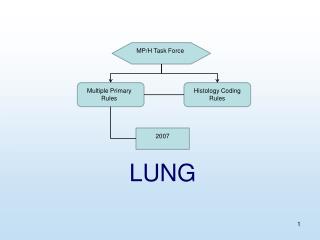
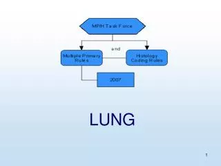
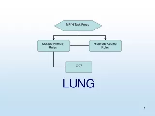
What?. Why?. 1. 2. -2.8kPa. -0.8kPa. TLC. FRC. - 2.2kPa. -0.3kPa. Coupling simulation of lung blood flow to pressure in the deforming lung tissue. K.S. Burrowes 1,2 & M.H. Tawhai 2.

E N D
What? Why? 1 2 -2.8kPa -0.8kPa TLC FRC -2.2kPa -0.3kPa Coupling simulation of lung blood flow to pressure in the deforming lung tissue K.S. Burrowes1,2 & M.H. Tawhai2 1 Dept. of Physiology & Computing Laboratory, University of Oxford, U.K. 2 Bioengineering Inst-itute, The University of Auckland, New Zealand Towards realistic predictions of pulmonary blood flow IUPS PHYSIOME PROJECT • We constructed a computational model to understand the interdependence between structure, fluid transport, and mechanical function in the lung. The use of a model provides exact control over functional parameters and the geometry of the solution domain. • The model of coupled soft-tissue mechanics-blood flow has been used to investigate flow heterogeneity in response to gravity, orientation of the lung, and resultant tissue deformation. This poster details the evolution towards a realistic model of pulmonary blood flow. 7 • Assumptions of a symmetric vascular network will produce unrealistic homogeneous flow distributions. • Coupling the flow model with a model of soft-tissue mechanics brings us even closer to making realistic predictions of blood flow distributions and enables calculations of flow at different lung volumes and in different postures , Fig 10. • We use a combination of medical imaging and a volume-filling branching algorithm to construct geometric models of the lung surface1 and vascular trees2, Fig 1. • Fig 10 compares inversion of postures: changes in lung volume produce a roughly linear reduction in flow from TLC to FRC. • Results demonstrate alterations in flow gradients resulting from inversion of posture, however flow gradients are not completely reversed indicating a component of structural dependence. Fig 1: Imaging-based model geometry. Spatially-consistent finite element models of lung, airway, and vascular trees derived from MDCT imaging in a single human subject. FLOW IN VARIOUS POSTURES • The use of an anatomically-based model enables more realistic flow predictions to be obtained. Fig 2 compares flows in the anatomically-based versus a symmetric branching model, illustrating that flow heterogeneity results from the asymmetric vascular branching structure. MODEL GEOMETRY Fig 10: (a) Terminal flows (30 mm slices) upright and inverted at TLC and FRC, height is cranio-caudal axis, and (b) prone and supine at TLC and FRC, height is dorso-ventral axis (0% at ventral surface). Fig 2: Flow solutions in anatomically-based (A) versus symmetric (S) model plotted with respect to height. 6 • Pressure fields computed from elastic deformation of the lung (Ppl) are coupled to the vascular pressure-radius relationship, Fig 8. • A reduced form of the Navier-Stokes equations and a pressure-radius relationship are solved within the vascular geometry to predict pressure, radius, and flow3. • The effect of Ppl and length changes on flow were assessed separately. Change in Ppl with constant geometry resulted in a relat-ively linear increase in flow from FRC to TLC due to increased radius values, Fig 9a. The flow response to vascular length deformations was more heterogeneous with higher average flows at FRC, Fig 9b. EVOLUTION: from symmetry to coupled mechanics-blood flow predictions • Diameters are allocated using a Strahler-based diameter ratio. Smaller diameter ratios result in vessels with larger radii therefore increased flow and a larger gradient of flow with respect to gravity, Fig 3. COUPLED MECHANICS-FLOW FLOW MODEL + GRAVITY (a) (b) (a) (b) Fig 3: (a) Radius and (b) flow (30 mm slices)versus height with various Strahler-based diameter ratios. Fig 8: Dependence of flow on mechanics solution in FRC lung upright: with mechanics, without mechanics (inlet Ppl=-5 cmH2O), with-out Ppl, & without Ppl or gravity. Height (%) refers to gravitationally-dependent height (i.e cranio-caudal axis). Fig 9: Flow averaged within 1 and 30 mm slices with respect to gravitationally-dependent height. (a) Within FRC geometry with TLC and FRC Ppl from mechanics solution. (b) FRC Ppl distribution from mechanics solution within FRC and TLC geometries calculated via mechanics model. Fig 4: Flow plotted with respect to height with various amounts of gravity averaged within 1 & 50 mm slices. • The effect of gravity on flow solutions was investigated, Fig 4, illustrating a clear influence of gravity but a consistent overall flow distribution. 3 4 5 SOFT-TISSUE MECHANICS • This study uses a model of the left human lung and arterial tree. The arterial model is embedded within the lung and deforms according to the change in lung volume, Fig 7. • Vessel elasticity has a significant effect on the distribution of flow, with more elastic vessels leading to a more pronounced gradient of flow due to gravity, Fig 5. • Lung volume changes are predicted using equations for finite deformation (large strain) theory, Fig 6, tissue pressure gradients are displayed in Table 1. Table 1: Slope of straight line fit to tissue pressures, as an estimate of Ppl gradient. • The material is described using a strain-energy density function6 with coefficients such that uniform inflation pressures are ~5cmH2O at FRC and ~25cmH2O at TLC. VESSEL ELASTICITY Posture Slope (cmH2O/cm) Upright TLC 0.220 (R2 =0.93) Upright FRC 0.174 (R2 =0.94) Inverted TLC -0.224 (R2 =0.91) Inverted FRC -0.164 (R2 =0.91) TLC VESSELS EMBEDDED FRC • The deformable lung model is contained by a contact ‘pleural’ body that changes geometry from TLC to FRC. Contact is enforced between the lung and the pleural surfaces, but the lung is free to slide. Figure 6: Surface pressures (kPa) and tissue deformation simulated in the upright lung at 1G, TLC and FRC. The hashed white surface and blue lines are on the contact body; coloured surface and red lines are the deformable lung. Fig 5: Variation in vessel elasticity (G0): Terminal flow values, averaged within 30 mm slices, plotted with respect to gravitationally-dependent height. G0 normally set as 5 kPa, based on experimental data4,5. Fig 7: Coupled mechanics-blood flow solution: Pressure distribution (kPa) at 1G within the upright left lung arterial model at TLC and FRC. • The reference geometry is taken as a uniform scaling from TLC to 25% of TLC. SUMMARY • Factors not yet accounted for: • Changes in the geometry of the chest wall, diaphragm, and heart with change in posture have not been included; • Coefficients in the material law were based on a simple assumption of Ppl(FRC)=-5cmH2O and Ppl(TLC)=-25cmH2O; • The effect of blood pressure was not included in the mechanics solutions; • Vessel elasticity did not change with changing length which would tend to decrease the flow predictions at TLC. ACKNOWLEDGEMENTS This work was supported by NIH grant R01-HL-064368, a Maurice Paykel Postdoctoral Fellowship and the Wellcome Trust in Oxford. The authors gratefully acknowledge the contributions of colleagues at the University of Iowa, Auckland, & Oxford. • This poster demonstrates steps towards producing a realistic model of blood flow in the lung. • Blood flow results display a relatively large amount of flow heterogeneity in all regions of the lung and a consistent decrease in flow in the cranial and caudal regions highlighting the significance of the vascular branching structure on flow distribution. • Coupling soft-tissue mechanics to blood flow increases understanding of the effect of lung tissue deformation on blood flow. Gravity has a more significant effect on tissue deformation at FRC than TLC, but a larger effect on blood flow at TLC than FRC due to greater distension of vessels. • REFERENCES • Tawhai et al, (2004), J Appl Physiol, 97(6):2310-2321. • Burrowes et al, (2005), J Appl Physiol ,99(2):731-738. • Burrowes et al, (2005), ‘Respir Physiol Neurobiol (in press). • Yen et al, (1980), J Biomech Eng 102: 170–177. • Krenz et al, (2003), Am J Physiol Heart Circ Physiol 284, H2192–H2203. • Fung, Y.C., (1975), Circ Res, 37: 481-496.