Download
1 / 68
680 likes | 702 Views
Explore the lymphatic system, blood volume protection, lymph vessels, lymphatic tissue, and human lymphoid organs. Learn about lymphocytes development, innate and adaptive immune responses, and mechanisms of immunological tolerance.

E N D
Lecture 12 The Lymphatic and Immune System
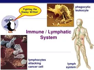
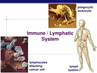
Lymphatic system Blood volume Protection Lymph and lymph vessels Lymphatic tissue Spleen Thymus
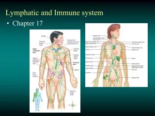
Human lymphoid organs Lymphocytes (thymus and bone marrow, which are therefore called central (or primary) lymphoid organs. The newly formed lymphocytes migrate from these primary organs to peripheral (or secondary) lymphoid organs, where they can react with foreign antigen. Only some of the peripheral lymphoid organs (blue) and lymphatic vessels (green) are shown; many lymphocytes, for example, are found in the skin and respiratory tract. Lymphatic vessels ultimately empty into bloodstream
The development of T and B cells The central lymphoid organs, where lymphocytes develop from common lymphoid progenitor cells, are labeled in yellow boxes. The common lymphoid progenitor cells develop from multipotent hemopoietic stem cells in the bone marrow. Some of the common lymphoid progenitor cells develop locally in the bone marrow into immature B cells, while others migrate to the thymus (via the bloodstream) where they develop into thymocytes (developing T cells). T cells and B cells are activated by foreign antigens mainly in peripheral lymphoid organs, such as lymph nodes or the spleen.
Innate and adaptive immune responses Innate immune responses are activated directly by pathogens and defend all multicellular organisms against infection. In vertebrates, pathogens, together with the innate immune responses they activate, stimulate adaptive immune responses, which then work together with innate immune responses to help fight the infection.
The two main classes of adaptive immune responses Lymphocytes carry out both classes of responses. Here, the lymphocytes are responding to a viral infection. In one class of adaptive response, B cells secrete antibodies that neutralize the virus. In the other, a T-cell-mediated response, T cells kill the virus-infected cells.
lymphocytes are required for adaptive immune responses to foreign antigens An important requirement of all such cell-transfer experiments is that cells are transferred between animals of the same inbred strain. Members of an inbred strain are genetically identical. If lymphocytes are transferred to a genetically different animal that has been irradiated, they react against the “foreign” antigens of the host and can kill the animal. In the experiment shown, the injection of lymphocytes restores both antibody and T-cell-mediated adaptive immune responses, indicating that lymphocytes are required for both types of responses.
How the innate immune system can help activate the adaptive immune system Dendritic cells ingest invading microbes. The microbial PAMPs activate the dendritic cells to express co-stimulatory proteins on their surface. The activated dendritic cells activate the T cells that express a receptor for the microbial antigens displayed on the dendritic cell surface. These T cells proliferate and some then migrate to the site of infection.
Electron micrographs of resting and effector lymphocytes This resting lymphocyte could be either a T cell or a B cell, as these cells are difficult to distinguish morphologically until they have been activated to become effector cells. (B) An effector B cell (a plasma cell). It is filled with an extensive rough endoplasmic reticulum (ER), which is distended with antibody molecules. (C) An effector T cell, which has relatively little rough ER but is filled with free ribosomes.
The dinitrophenyl (DNP) group Although it is too small to induce an immune response on its own, when it is coupled covalently to a lysine side chain on a protein, as illustrated, DNP stimulates production of the hundreds of different species of antibodies that all bind specifically to it
A model for the cellular basis of immunological memory When stimulated by their specific antigen, naïve cells proliferate and differentiate. Most become effector cells, which function and then usually die, while others become memory cells. During a subsequent exposure to the same antigen, the memory cells respond more readily, rapidly, and efficiently than did the naïve cells: they proliferate and give rise to effector cells and to more memory cells.
Acquired immunological tolerance The skin graft seen here was transplanted from an adult brown mouse to an adult white mouse. It has survived for many weeks only because the white mouse, at the time of its birth, received an injection of bone marrow cells from the brown mouse and therefore became immunologically tolerant to them. Some of the bone marrow cells (and their progeny) from the brown mouse persist in the adult white mouse and continue to induce tolerance in newly formed lymphocytes that would otherwise react against the brown skin.
Mechanisms of immunological tolerance to self antigens 1. In receptor editing, developing lymphocytes that recognize self molecules (self-reactive lymphocytes) change their antigen receptors so that they no longer recognize self antigens. 2. In clonal deletion, self-reactive lymphocytes die by apoptosis when they bind their self antigen. 3. In clonal inactivation (also called clonal anergy), self-reactive lymphocytes become functionally inactivated when they encounter their self antigen. 4. In clonal suppression, regulatory T cells suppress the activity of self-reactive lymphocytes.
The path followed by lymphocytes as they continuously circulate between the lymph and blood The circulation through a lymph node (yellow) is shown here. Microbial antigens are usually carried into the lymph node by dendritic cells (not shown), which enter the node via afferent lymphatic vessels draining an infected tissue (green). T and B cells, by contrast, enter the lymph node via an artery and migrate out of the bloodstream through postcapillary venules. Unless they encounter their antigen, the T and B cells leave the lymph node via efferent lymphatic vessels, which eventually join the thoracic duct. The thoracic duct empties into a large vein carrying blood to the heart to complete the circulation process for T and B cells. A typical circulation cycle for these lymphocytes takes about 12–24 hours.
Migration of a lymphocyte out of the bloodstream into a lymph node A circulating lymphocyte adheres weakly to the surface of the specialized endothelial cells by L-selectin on the lymphocyte surface. Stimulated by chemokines secreted by the endothelial cells (curved red arrow), the lymphocyte rapidly activates a stronger adhesion system, mediated by an integrin. This strong adhesion enables the cell to stop rolling. The lymphocyte then uses an Ig like cell adhesion protein (CD31) to bind to the junctions between adjacent endothelial cells and migrate out of the venule. CD31 is located both on the surface of the lymphocyte and at the junctions between the endothelial cells. The subsequent migration of the lymphocytes in the lymph node depends on chemokines produced within the node (straight red arrow).
A simplified drawing of a human lymph node B cells are primarily clustered in structures called lymphoid follicles, whereas T cells are found mainly in the paracortex. Chemokines attract both types of lymphocytes into the lymph node from the blood via postcapillary venules. T and B cells then migrate to their respective areas, attracted by different chemokines. If they do not encounter their specific antigen, both T cells and B cells then enter the medullary sinuses and leave the node via the efferent lymphatic vessel. This vessel ultimately empties into the bloodstream, allowing the lymphocytes to begin another cycle of circulation through a peripheral lymphoid organ. If they encounter their specific antigen, T and B cells are retained in the node and are activated to become effector cells or memory cells; T cells and B cells responding to the same pathogen can interact in and around lymphoid follicles
The membrane-bound and secreted antibodies made by a B cell clone When an antigen (aided by a helper T cell—not shown) binds to and thereby activates either a naïve or a memory B cell, the cell proliferates and differentiates into effector cells. The effector cells produce and secrete antibodies with a unique antigen-binding site, which is the same as that of their original membrane-bound antibody that served as their antigen receptors.
Antibody–antigen interactions Because antibodies have two identical antigen-binding sites, they can cross-link antigens. The types of antibody–antigen complexes that form depend on the number of antigenic determinants on the antigen. (A–C) A single species of antibody (a monoclonal antibody) is shown binding to antigens containing one, two, or three copies of a single type of antigenic determinant. Antigens with two identical antigenic determinants can form small cyclic complexes or linear chains with the antibodies, while antigens with three or more identical antigenic determinants can form large three-dimensional lattices that readily precipitate out of solution. (D) Most antigens have many different antigenic determinants, and different antibodies that recognize different determinants can cooperate in cross-linking the antigen into large three-dimensional lattices.
The hinge region of an antibody molecule Because of its flexibility, the hinge region improves the efficiency of antigen binding and crosslinking
A schematic drawing of a bivalent antibody molecule It is composed of four polypeptide chains—two identical heavy chains and two identical light chains. The two antigen binding sites are identical, each formed by the N-terminal region of a light chain and the N-terminal region of a heavy chain. The two heavy chains also form both the tail and hinge region of the antibody.
The main stages in B cell development All of the stages shown occur independently of antigen. The pro-B cell makes m chains, but they remain in the endoplasmic reticulum until surrogate light chains are made. Although not shown, all of the cell-surface Ig molecules are associated with transmembrane proteins that help convey signals to the cell interior. When they are activated by their specific foreign antigen and helper T cells in peripheral lymphoid organs, mature naïve B cells proliferate and differentiate into either antibody-secreting cells or memory cells (not shown).
A pentameric IgM molecule Disulfide bonds (red) hold the five four-chain units together. A single J chain, which has a structure similar to that of a single Ig domain is covalently attached by disulfide bonds to the tails of two m heavy chains. The J chain is required for pentamer formation. The addition of each successive four-chain IgM unit requires a J chain, which is then discarded, except for the last one, which is retained. Note that IgM molecules do not have hinge regions.
A highly schematized diagram of a dimeric IgA molecule found in secretions In addition to the two IgA monomers, there is a single J chain and an additional polypeptide chain called the secretory component, which is derived from the Fc receptor and is thought to protect the IgA molecules from digestion by proteolytic enzymes in secretions.
The mechanism of transport of a dimeric IgA molecule across an epithelial cell The IgA molecule, as a J-chain-containing dimer, binds to a transmembrane receptor protein on the nonluminal surface of a secretory epithelial cell. The receptor–IgA complexes are ingested by receptor mediated endocytosis, transferred across the epithelial cell cytoplasm in vesicles, and secreted into the lumen on the opposite side of the cell by exocytosis. When exposed to the lumen, the part of the Fc receptor protein that is bound to the IgA dimer (the secretory component) is cleaved from its transmembrane tail, thereby releasing the antibody.
The role of IgE in histamine secretion by mast cells A mast cell (or a basophil) binds IgE molecules after they are secreted by effector B cells. The soluble IgE antibodies bind to Fc receptor proteins on the mast cell surface that specifically recognize the Fc region of these antibodies. The bound IgE molecules serve as cell-surface receptors for antigen. Thus, unlike B cells, each mast cell (and basophil) has a set of cell-surface antibodies with a wide variety of antigen-binding sites. When an antigen molecule binds to these membrane-bound IgE antibodies so as to cross-link them to their neighbors, it signals the mast cell to release its histamine and other local mediators by exocytosis.
Antigen binding to antibody In this diagram, an antigenic determinant on a macromolecule is shown interacting with one of the antigen-binding sites of two different antibody molecules, one of high affinity and one of low affinity. Various weak noncovalent forces hold the antigenic determinant in the binding site, and the site with the better fit to the antigen has a greater affinity. Note that both the light and heavy chains of the antibody molecule usually contribute to the antigen-binding site.
Molecules with multiple antigenic determinants (A) A globular protein is shown with a number of different antigenic determinants. Different regions of a polypeptide chain usually come together in the folded structure to form each antigenic determinant on the surface of the protein, as shown for three of the four determinants. (B) A polymeric structure is shown with many identical antigenic determinants.
Constant and variable regions of immunoglobulin chains The variable regions of the light and heavy chains form the antigen-binding sites, while the constant regions of the heavy chains determine the other biological properties of an antibody.
Antibody hypervariable regions Highly schematized drawing of how the three hypervariable regions in each light and heavy chain together form the antigen-binding site of an antibody molecule.
The variety of antigen binding surfaces in antibodies The hypervariable loops of different VL and VH domains can combine to form a large variety of binding surfaces. The antigenic determinants and the antigen-binding site of the antibodies are shown in red. Only one antigen-binding site is shown for each antibody.
Three types of proteins on the surface of an activated dendritic cell involved in activating a T cell. The invariant polypeptide chains that are always stably associated with the T cell receptor (TCR) are not shown
Effector cytotoxic T cells killing target cells in culture. (A) Electron micrograph showing an effector cytotoxic T cell binding to a target cell. The cytotoxic T cells were obtained from mice immunized with the target cells, which are foreign tumor cells. (B) Electron micrograph showing a cytotoxic T cell and a tumor cell that the T cell has killed. In an animal, as opposed to in a culture dish, the killed target cell would be phagocytosed by neighboring cells long before it disintegrated in the way that it has here. (C) Immuno-fluorescence micrograph of a T cell and tumor cell after staining with anti-tubulin antibodies. Note that the centrosome in the T cell is located at the point of cell–cell contact with the target cell—an immunological synapse. The secretory granules (not visible) in the T cell are initially transported along microtubules to the centrosome, which then moves to the synapse, delivering the granules to where they can release their contents.
Two strategies by which effector cytotoxic T cells kill their target cells
Differentiation of naïve helper T cells into either TH1 or TH2 effector helper cells in a peripheral lymphoid organ The nature of the activated dendritic cell and the characteristics of the pathogen that activated it mainly determine which type of effector helper cell develops.
Recognition by T cells of foreign peptides bound to MHC proteins Cytotoxic T cells recognize foreign peptides in association with class I MHC proteins, whereas helper T cells and regulatory T cells recognize foreign peptides in association with class II MHC proteins. In both cases, the T cell recognizes the peptide–MHC complexes on the surface of a dendritic cell or a target cell.
Human MHC gene This simplified schematic drawing shows the location of the genes that encode the transmembrane subunits of class I (light green) and class II (dark green) MHC proteins. The genes shown encode three types of class I proteins (HLA-A, HLA-B, and HLA-C) and three types of class II MHC proteins (HLA-DP, HLA-DQ, and HLA-DR). An individual can therefore make six types of class I MHC proteins and more than six types of class II MHC proteins.
The three-dimensional structure of a human class I MHC protein as determined by x-ray diffraction analysis of crystals of the extracellular part of the molecule
The interaction of a T cell receptor with a viral peptide bound to a class I MHC protein
CD4 and CD8 co-receptors on the surface of T cells Cytotoxic T cells (TC) express CD8, which recognizes class I MHC proteins, whereas helper T cells (TH) and regulatory T cells (not shown) express CD4, which recognizes class II MHC proteins. Note that the co-receptors bind to the same MHC protein that the TCR has engaged, so that they are brought together with TCRs during the antigen recognition process. Whereas the TCR binds to the variable (polymorphic) parts of the MHC protein that form the peptide-binding groove, the co-receptor binds to the invariant part, well away from the groove.
The classic experiment showing that an effector cytotoxic T cell recognizes some aspect of the surface of the host target cell in addition to a viral antigen
The peptide-transport problem How do peptide fragments get from the cytosol, where they are produced, into the ER lumen, where the peptide-binding grooves of class I MHC proteins are located? A special transport process is required.