Download
1 / 42
560 likes | 1.39k Views
CT. Seeram Chapter 9: Image Manipulation in CT. Image Manipulation Defined. “Those techniques (operations) or processes which modify an image or group of images to enhance the visibility of useful information while suppressing ‘noise’ or non-useful information”

E N D
CT Seeram Chapter 9: Image Manipulation in CT
Image Manipulation Defined • “Those techniques (operations) or processes which modify an image or group of images to enhance the visibility of useful information while suppressing ‘noise’ or non-useful information” • No additional or new information produced • Content of interest emphasized • Overall information content reduced
Windowing • Manipulation of image gray scale using image’s CT numbers
Windowing • Manipulation allows customization of visibility • soft tissues • brain • dense structures • bone
Window Width & Level • Window width • range of CT #’s imaged • determines maximum # of gray shades which could be displayed on monitor • Window level • center or midpoint of CT # range WL WW
Window Width & Level • Pixels outside of window displayed as Black or White WL WW
Window Width & Level 3000 >200 151-200 2000 101-150 51-100 1-50 1000 (-49)-0 (-99)-(-50) Window: 400 Level: 0 (-149)-(-100) 0 (-199)-(-150) <(-199) -1000
1000 Small Window Width 3000 • Short gray scale • Small block of CT #’s assigned gray levels • Small transition zone of white to black Window: 400 Level: 0 0 -1000 -1000
1000 Small Window Width 3000 • Used to display soft tissues within structures containing different tissues of similar densities • Level centered near average CT # of organ of interest Window: 400 Level: 0 0 -1000 -1000
1000 Large Window Width • Long gray scale • Large block of CT #’s assigned gray levels • Large transition zone of white to black Window: 2000 Level: 0 0 -1000
1000 Large Window Width • Used where large latitude required • Used to simultaneously display tissues of greatly differing attenuation Window: 2000 Level: 0 0 -1000
Window Example 100 WL =0 WW = 200 All pixels with CT #’s > 0 +(200/2) = 100: White 0 200 All pixels with CT #’s < 0 -(200/2) = -100: Black -100
Another Window Example 140 WL = 40 WW = 200 All pixels with CT #’s > 40 + (200/2) = 140: White 40 200 All pixels with CT #’s < 40 - (200/2) = -60: Black -60
Still Another Window Example 200 WL = 0 WW = 400 All pixels with CT #’s > 0 + (400/2) = 200: White 0 400 All pixels with CT #’s < 0 - (400/2) = -200: Black -200
Larger Window Means Obscuring Small Differences in Tissue Attenuation WW=200 WW=400 200 100 • One gray shade encompasses larger range of CT #’s Range 20 - 40 40 - 80 20 40 -100 -200
Window Width & Contrast • As WW increases • contrast decreases • latitude (range of CT #’s imaged) increases • As WW decreases • contrast increases • latitude decreases • Clinical goal: • Largest available contrast at the latitude required by study
Window Width & Image Contrast • Large window width • different structures more likely to have same gray shade • Narrow window width • Gray shade differences more likely visible between structures • Very narrow window width • Small differences in attenuation seen as black & white
Preset Window & Level • Available for all commercial CT • initial WW and WL pre-sets for specific study types • Can be overridden by operator
Specialized CT Image Manipulation Programs • Region of interest (ROI) analysis • average CT # • standard deviation
Specialized CT Image Manipulation Programs • Image annotation • grids • arrows • notes Bad Thingee
Specialized CT Image Manipulation Programs • Histogram analysis • graph of CT # frequency • # of pixels with given range of CT#’s • can apply to • ROI • entire image
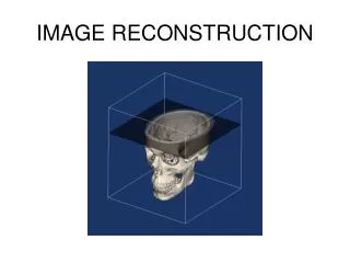
Specialized CT Image Manipulation Programs • Multiplanar reconstruction • 3D reconstruction • Quantitative CT • Osteo CT / bone densitometry • Xenon CT • Radiation therapy treatment planning using CT data
Multiplanar Reconstruction • Creates non-axial images from stack of contiguous transverse axial scans • stack contains 3 dimensional CT data • pixels new cut identified & selected from each axial image without scanning
Cuts • coronal • sagittal • paraxial • oblique
Coronal Reconstruction Example Stack of Axial Slices Reconstructed Slice
Coronal Reconstruction Example Reconstructed Slice Stack of Axial Slices
Sagittal Reconstruction Example Reconstructed Slice Stack of Axial Slices
Oblique Reconstruction Example Reconstructed Slice Stack of Axial Slices
Reformatting Advantages • enables visualization of specific structures relative to surrounding structures • aids in determining / localizing true extent of • lesions • fractures • bone fragments • foreign bodies
Reformatting Disadvantages • Image quality can be poorer than axial images if plane thickness > pixel size • affects blurring • less problem in spiral scanning
Reformatting Disadvantages • More prone to motion / breathing artifacts • reformatted image taken from many slices • reformatted image represents longer time interval than single slice
Quantitative CT: Measurement of Bone Mineral Density (BMD) • Competes with DEXA
Quantitative CT: Measurement of Bone Mineral Density (BMD) • Scout image used to define midvertebral plane • Axial images contains both patient & reference phantom • Reference phantom used to account for slight CT # changes over time • phantom has water & bone equivalent parts
Quantitative CT: Measurement of Bone Mineral Density (BMD) • Software automatically defines region of interest • Mean CT #’s calculated • Bone Mineral Density calculated • BMD compared to • “standard” values • previous studies for this patient
Xenon CT for Regional Cerebral Blood Flow Imaging • Patient inhales xenon gas • serves as a contrast agent • Xenon • gas • inert element • high atomic # • provides good CT contrast • high fat solubility • crosses blood - brain barrier
Xenon CT Applications • Brain metabolism • regional perfusion in cerebrovascular disease • Potential applications • dementia • sleep disorders • migraines • epilepsy
CT in Radiation Therapy Treatment Planning • Conventional CT study performed • patient positioned exactly as for therapy • CT data communicated to treatment planning computer
CT in Radiation Therapy Treatment Planning • CT data used to develop depth dose data • Isodose data superimposed on CT image
Other CT Software Features • Multiple window settings within a slice • Image magnification • Measurement of distances / angles • Highlighting of selected pixel values in image • Cine viewing of image stack
CT Applications • Split image • one thicker slice reconstructed into two thinner slices • Dental • Angiography • Plastic Surgery Reconstruction • Multimodality Image Fusion • Overlaying of images of various modalities • One anatomical (CT), one functional (Nuc med)