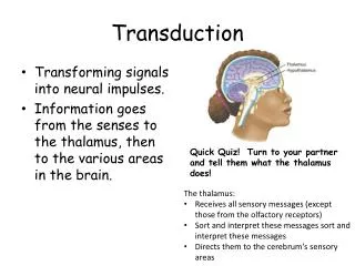
Visual Transduction
Visual Transduction. Once light waves have been successfully focused on the retina, the information “stored” in that electromagnetic energy must be changed by photopigments in the photoreceptors into signals our brain can interpret - a process called visual transduction

Visual Transduction
E N D
Presentation Transcript
Visual Transduction • Once light waves have been successfully focused on the retina, the information “stored” in that electromagnetic energy must be changed by photopigments in the photoreceptors into signals our brain can interpret - a process called visual transduction • The single type of photopigment in rods is rhodopsin, whereas there are 3 different cone photopigments • Color vision results from different colors of light selectively activating the different cone photopigments
Visual Transduction • The first step in visual transduction is absorption of light by a photopigment, a colored protein that undergoes structural changes when it absorbs light in the outer segment of a photoreceptor • Light absorption initiates a series of events that lead to the production of a receptor potential (number 4 in the diagram)
Visual Transduction • All photopigments associated with vision contain two parts: a glycoprotein known as opsinand a derivative of vitamin A called retinal • Although there are 4 different opsins, retinal is the light-absorbing part of all visual photopigments • To simplify the process we can say that there is a cyclical bleaching and regeneration of photopigment • Bleaching is a term describing a conformational change in the retinal molecule in response to light
Visual Transduction • In darkness, retinal has a bent shape called cis-retinal • Absorption of a photon of light causes it to straighten into the trans-retinal form in a process called isomerization • Trans-retinal completely separates from the opsin; since the final products look colorless, this part of the cycle is called bleaching of photopigment • An enzyme converts trans-retinal→ cis-retinal • The cis-retinal regenerates the photopigment
Bleaching and regeneration of photopigments are summarized here
Visual Transduction • In daylight, regeneration of rhodopsin cannot keep up with the bleaching process, so rods contribute little to daylight vision. In contrast, cone photopigments regenerate rapidly enough that some of the cis form is always present, even in very bright light • As a consequence, light adaptation (from dark conditions light conditions) happens in seconds; dark adaptation (from light dark) takes minutes to occur (up to 40 minutes to fully adapt)
Visual Transduction • Most forms of color blindness, an inherited inability to distinguish between certain colors, result from the absence or deficiency of one of the three types of cones • Most common type is red-green color blindness in which red cones or green cones are missing • Prolonged vitamin A deficiency and the resulting below-normal amount of rhodopsin may cause night blindness or nyctalopia, an inability to see well at low light levels
The Visual Pathway • The graded potentials generated by the photoreceptors undergo considerable processing at synapses among the various types of neurons in the retina (horizontal cells, bipolar cells, and amacrine cells)- certain features of visual input are enhanced while others are discarded • Overall, convergence pre- dominates as 126 million photo-receptors impinge on only 1 million ganglion cells
The Visual Pathway • The axons of retinal ganglion cells provide output that travels back “towards the light”, exiting the eyeball as the optic nerve,whichemerges from the vitreous surface of the retina • The axons then pass through a crossover point called the optic chiasm
The Visual Pathway • Some axons cross to the opposite side, while others remain uncrossed. Once through the optic chiasm the axons enter the brain matter as the optic tracts (most terminate in thalamus) • Here they synapse with neurons that project to the 1o visual cortex in the occipital lobes
Visual field of left eye Visual field of left eye Visual field of left eye Visual field of left eye Visual field of left eye Visual field of left eye Visual field of right eye Visual field of right eye Visual field of right eye Visual field of right eye Visual field of right eye Visual field of right eye Temporal half Temporal half Temporal half Temporal half Temporal half Temporal half Nasal half Nasal half Nasal half Nasal half Nasal half Nasal half Nasal half Nasal half Nasal half Nasal half Nasal half Nasal half Temporal half Temporal half Temporal half Temporal half Temporal half Temporal half Left eye Left eye Left eye Left eye Left eye Left eye Right eye Right eye Right eye Right eye Right eye Right eye Nasal retina Nasal retina Nasal retina Nasal retina Nasal retina Nasal retina Temporal retina Temporal retina Temporal retina Temporal retina Temporal retina Temporal retina Nasal retina Nasal retina Nasal retina Nasal retina Nasal retina Nasal retina Temporal retina Temporal retina Temporal retina Temporal retina Temporal retina Temporal retina 1 1 1 1 1 1 3 3 3 3 1 1 1 1 1 1 3 3 3 3 4 4 4 4 4 4 Optic tract Optic tract Optic tract 2 2 2 2 2 2 2 2 2 2 Midbrain Midbrain Midbrain Midbrain Midbrain Midbrain Midbrain Midbrain Midbrain Midbrain Midbrain Midbrain 5 5 5 5 Lateral geniculate nucleus of the thalamus Lateral geniculate nucleus of the thalamus Lateral geniculate nucleus of the thalamus Lateral geniculate nucleus of the thalamus Lateral geniculate nucleus of the thalamus Lateral geniculate nucleus of the thalamus 6 Optic radiations Optic radiations Optic radiations Optic radiations Optic radiations Optic radiations Optic radiations Optic radiations Optic radiations Optic radiations Optic radiations Optic radiations 6 Primary visual area of cerebral cortex (area 17) in occipital lobe Primary visual area of cerebral cortex (area 17) in occipital lobe Primary visual area of cerebral cortex (area 17) in occipital lobe Primary visual area of cerebral cortex (area 17) in occipital lobe Primary visual area of cerebral cortex (area 17) in occipital lobe Primary visual area of cerebral cortex (area 17) in occipital lobe Left eye and its pathways Left eye and its pathways Left eye and its pathways Left eye and its pathways Left eye and its pathways Left eye and its pathways Right eye and its pathways Right eye and its pathways Right eye and its pathways Right eye and its pathways Right eye and its pathways Right eye and its pathways
The Ear • Audition, the process of hearing, is accomplished by the organs of the ear. The ear is an engineering marvel because its sensory receptors can transduce sound vibrations with amplitudes as small as the diameter of an atom of gold into electrical signals 1000 times faster than the eye can respond to light • The ear also contains receptors for equilibrium
The Ear • The ear has 3 principle regions • The external ear, which uses air to collect and channel sound waves • The middle ear, which uses a bony system to amplify sound vibrations • The internal ear, which generates action potentials to transmit sound and balance information to the brain
The External Ear • The anatomy of the external ear includes • The auricle (pinna), a flap of elastic cartilage covered by skin and containing ceruminous glands • A curved 1” long external auditory canal situated in the temporal bone leading from the meatus to the tympanic membrane (TM – or ear drum) which separates the outer ear from the cavity of the middle ear
The Middle Ear • The middle ear is an air-filled cavity in the temporal bone. It is lined with epithelium and contains 3 auditory ossicles (bones) • The stapes (stirrup) • The incus (anvil) • The handle of the malleus (hammer) attaches to the TM
The Middle Ear • Two small skeletal muscles (the tensor tympani and stapedius) attach to the ossicle and dampen vibrations to prevent damage from sudden, loud sounds
The Middle Ear • The Eustachian (auditory) tube connects the middle ear with the nasopharynx (upper portion of the throat) • It consists of bone and hyaline cartilage and is normally passively collapsed. It opens to equalize pressures on each side of the TM (allowing it to vibrate freely)
The Inner Ear • The internal ear (inner ear) is also called the labyrinth because of its complicated series of canals • Structurally, it consists of two main divisions: an outerbony labyrinththat encloses an inner membranous labyrinth • the bony labyrinth is sculpted out of the petrous part of the temporal bone, and divided into three areas: (1) the semicircular canals, (2) the vestibule, and (3) the cochlea
The Inner Ear • The vestibule is the middle part of the bony labyrinth • The membranous labyrinth in the vestibule consists of two sacs called the utricle and the saccule (both contain rc for static equilibrium) • The cochlea , located anterior to the vestibule, contains rc for hearing • The three semicircular canals are above the vestibule, each ending in a swollen enlargement called the ampulla (for dynamic equilibrium)
The Inner Ear • The snail shaped cochlea contains the hearing apparatus • Two types of fluid (perilymph and endolymph) fill its 3 different internal channels: The scala vestibuli, scala tympani, and cochlear duct • A section through one turn of the cochlea is shown
The Inner Ear • Perilymph transmits the vibrations coming from the stapes in the oval window up and around the scala vestibuli, and then back down and around the scala tympani – causing the endolymph in the cochlear duct to vibrate • Pressure waves in the endolymph cause the basilar membrane of the cochlear duct to vibrate, moving the hair cells of the spiral organ of Corti against an overhanging flexible gelatinous membrane called the tectorial membrane
The Inner Ear • Note how the sound waves between the number 1 and number 2 in this diagram are shown impacting different parts of membranous labyrinth. This is a representation of sounds waves of different frequencies being transduced at the segment of the basilar membrane that is “tuned” for a particular pitch
The Inner Ear • Movements of the hair cells in contact with the tectorial membrane transduce mechanical vibrations into electrical signals which generate nerve impulses along the cochlear branch of CN VIII
The Auditory Pathway • This graphic depicts the events in the stimulation of auditory receptors, from channeling sound waves into the external ear and onto the TM, to the transduction of those vibrations into local receptor potentials