Setting Anatomic Posterior Teeth
170 likes | 481 Views
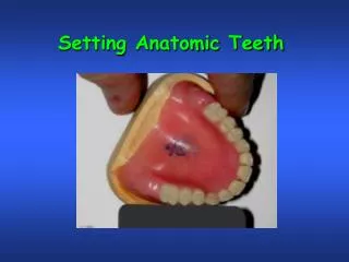
Setting Anatomic Posterior Teeth. These are the molds of teeth you should be using in this course (32M post., 4N max. ant., 2N mand. ant.). .

Setting Anatomic Posterior Teeth
E N D
Presentation Transcript
These are the molds of teeth you should be using in this course (32M post., 4N max. ant., 2N mand. ant.).
Remove a section of wax from one side of the maxillary baseplate between the canine and the second molar position. Leave a posterior pillar of wax in the 2nd molar area. A quick look will verify that the maxillary posterior teeth should have plenty of space to be set with little or no reduction. Reduce the baseplate if necessary, and if that is still not enough, reduce the ridgelap area of the teeth. Set the first premolar in proper buccolingual position.
The rim is left intact on the opposite side because this will help you to maintain the location of the occlusal plane. The pillar of wax that is left on the side where the teeth are to be set preserves the occlusal plane in this area and facilitates the setting of the 1st premolar and 1st molar on this plane. The posterior teeth are first set flat to the occlusal plane and with their central grooves aligned over the mark on the center of the mandibular ridge.
Once these two teeth are set, the pillar is removed and the 2nd molar is set to this plane that is preserved by these teeth and the wax rim on the opposite side. The second molar is set flat with the plane and along the same plane buccally that was established with the position of the 1st molar. A tongue blade is used to ensure that the 2nd molar lies along this buccal plane.
A tongue blade is used to verify that the buccal cusp of the maxillary premolar and the mesiobuccal cusp of the first molar lie along a plane with the middle of the facial surface of the Canine. A tongue blade is also used to verify that the buccal cusps of the1st and 2nd molars lie along a plane that is defined by turning the distobuccal cusp of the 1st molar 20o toward the palate.
The setup is checked from the posterior to ensure that the central groove of the 2nd molar is exactly over the crest of the mandibular ridge as defined by the mark on the mandibular occlusal rim.
A tongue blade is then used to depress the distobuccal cusp of the 1st molar so that it lies ½ mm above the plane. The same procedure is used to depress the buccal cusps of the 2nd molar so that they lie along this same plane, and that the mesiobuccal cusp is 1 mm above the occlusal plane and the distobuccal cusp is 1½ mm above the plane. The lingual cusps will be left extending down toward the occlusal plane.
When the plane is done properly, the maxillary 1st molar will provide the guide for the occlusal surface of the 2nd molar. All cusps of both molars should contact this plane.
Check this relationship with the opposing occlusion rim and the flat plate. There should be a noticeable rise in the occlusal plane from the distobuccal of the first molar through the second molar. All other teeth should be touching the plate except for the maxillary lateral incisor.
Anterior teeth with anatomic posterior teeth are set to have a vertical overlap equal to the compensating curve.
After the maxillary teeth are adjusted to provide the compensating curve, the buccolingual position of each posterior tooth is rechecked to ensure that it is still correct, as the shrinkage of the wax as it sets and contact of the teeth with the baseplate will have an effect on the position that a tooth can be stabilized. The mesiobuccal cusp of the 1st molar is in contact with a plane that runs to the midbuccal of the canine, with the premolar also in contact with this plane.
? ? ? ? ? ? ? Questions? Questions? Questions?