Download
1 / 39
410 likes | 611 Views
Overview of Cells and Cell Research. BL 424 Cell Biology Ch 1 Overview Student learning outcomes: 1. Describe Origin and Evolution of Cells – prokaryote and eukaryote 2. Explain Cells as Experimental Models – examples of model organisms

E N D
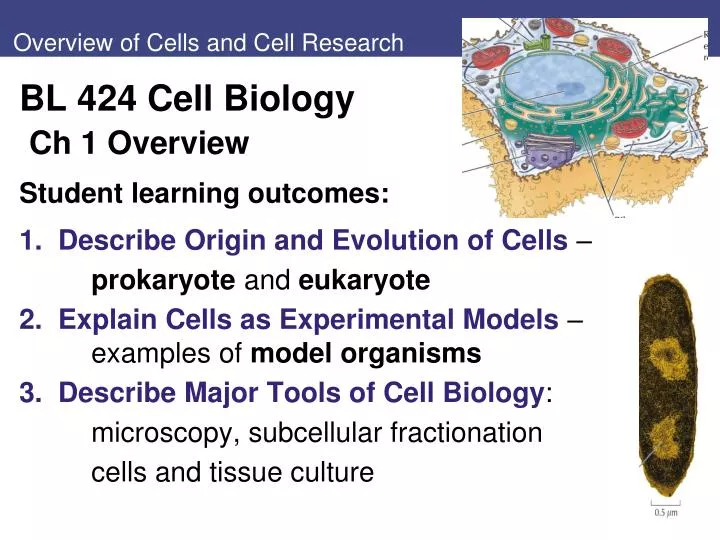
Overview of Cells and Cell Research • BL 424 Cell Biology • Ch 1 Overview • Student learning outcomes: • 1. Describe Origin and Evolution of Cells – • prokaryote and eukaryote • 2. Explain Cells as Experimental Models – examples of model organisms • 3. Describe Major Tools of Cell Biology: • microscopy, subcellular fractionation • cells and tissue culture
Unity and diversity of present-day cells reflects evolution from common ancestor: • Prokaryotic cells (Bacteria and Archaea): • lack nuclear envelope • Eukaryotic cells (yeast, plants, animals): • nuclear membrane encloses genetic material
Fig 1.3 Enclosure of self-replicating RNA in a phospholipid membrane Phospholipids: basic components of present-day biological membranes. RNA world theory: first cell arose from self-replicating RNA molecule in membrane Fig. 1.3 Phospholipids are amphipathic: Water-insoluble (hydrophobic) hydrocarbon chains Water-soluble (hydrophilic) head groups with PO4 In water, phospholipids spontaneously aggregate into bilayers.
The Origin and Evolution of Cells Adenosine 5′-triphosphate (ATP) metabolic energy 3 Mechanisms of generation of ATP: Glycolysis Photo- synthesis Oxidative metabolism Fig. 1.4
The Origin and Evolution of Cells Present-day prokaryotes: Archaea (Archaebacteria): many live in extreme environments Halobacterium; Methanooccus Bacteria(Eubacteria): large group - many environments: Escherichia, Bacillus Cyanobacteria, photosynthesis evolved; largest and most complex prokaryotes: Anabaena Fig. 1.5 E. coli : cell wall, plasma membrane, circular DNA without nuclear membrane
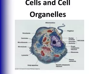
The Origin and Evolution of Cells Eukaryotic cells are larger, more complex: Plasma membrane, ribosomes Nucleus is large organelle: linear DNA molecules Membrane-bound organelles - specialized structures and functions Fig. 1.6: animal cell, plant cell
Fig 1.7 Evolution of cells • Endosymbiosis: eukaryote organelles arose from prokaryotic cells living inside ancestors of eukaryotes. • evidence strongest for mitochondria and chloroplasts • Mosaic nature of • eukaryotic genomes: • fusion of • archaeal and • eubacterial • genomes. • Archaea more closely • related to Eukarya Fig. 1.7
The Origin and Evolution of Cells Unicellular eukaryotes: Yeastis simple eukaryote Saccharomyces cerevisiae ~ 6 µm diameter; 12x106 bp DNA Protozoan Plasmodium falciparium causes malaria Gametocyte and blood cells
The Origin and Evolution of Cells Multicellular organisms - diversity of cell types for specialized functions: Animal cell tissue types: Epithelial cells - cover surfaces, line organs. 2. Connective tissues - bone, cartilage, adipose tissue. Loose connective tissue is formed by fibroblasts. 3. Blood cells: Red cells (erythrocytes) transport oxygen. White blood cells inflammatory, immune response. 4. Nervous tissue – neurons, supporting cells, sensory cells. Muscle cells Fig. 1.12 cells A, epithelial B, fibroblasts C, blood cells
2. Cells as Experimental Models 1.2 Cells as Experimental Models: Evolution conserved fundamental properties Basic principles from experiments on one type of cell generally applicable to other cells Some cells and organisms are good experimental models – model organisms. Unity of molecular cell biology: General principles of cell structure, function from studies of yeast apply to all eukaryotic cells. Understanding development of multicellular organisms requires analysis of plants, animals (Table 2 shows organisms vary widely in DNA content, genes)
Cells as Experimental Models Escherichia coli – E. coli Bacteria Basic features of DNA replication, genetic code, gene expression, and protein synthesis Genome only 4.6x10⁶ bp, 4300 genes Ease of culture in laboratory, Elucidate biochemical pathways; Easy to select genetic variants Can transfer partial genome to others Recombinant DNA, plasmids Divides every 20 min -> colony of 108 cells overnight Fig. 1.13
Cells as Experimental Models Yeastis simple eukaryote, model for fundamental studies of eukaryotic biology. Genome of Saccharomyces cerevisiae is 12x106 bp of DNA, about 6000 genes. Easily grown as colonies from single cell – grow as haploid or diploid Recombinant DNA techniques Two mating types Has cell wall Genetic manipulations similar to those performed using bacteria Fig. 1.14
Cells as Experimental Models – C. elegans NematodeCaenorhabditis elegans is widely used model: Fig. 1.15 Genome ~ 19,000 genes Adult worm only 959 somatic cells. Embryonic origin and lineage of all cells has been traced; grows fast Mutants with developmental abnormalities
Cells as Experimental Models – Drosophila, Arabidopsis Developmental biology: fruitfly, mouse-ear cress Drosophila melanogaster Arabidopsis thaliana Small genomes; short reproductive cycles; easy to find mutants Figs. 1.16, 1.17
Cells as Experimental Models - vertebrates Vertebrates - most complex animals, most difficult to study from cell, molecular biology. One approach uses cells in culture: culture cells in chemically defined media Highly differentiated cells are important models for particular aspects of cell biology. Ex. Muscle cells model for cell movement at molecular level. Ex. Giant neurons to study ion transport, cytoskeleton function.
Cells as Experimental Models – Xenopus, Danio Models for vertebrate development include: FrogXenopus laevis and Zebrafish(Danio rerio) Frog - large eggs in large numbers facilitates biochemical analysis Zebrafish are small, reproduce rapidly. Transparent embryos develop outside of mother; early stages of development can be easily observed. African clawed frog Figs. 1.18, 1.19
Cells as Experimental Models – Mus musculus Mouse(Mus musculus) is mammal model. Genetically engineer mice with specific mutant genes to study functions of genes, development Similarity of genomes: Mutations in homologous genes result in similar developmental defects Ex. Piebald mutation of gene for melanocyte migration Fig. 1.20
1.3 Tools of Cell Biology 1.3 Tools of Cell Biology: Research depends on available laboratory methods and experimental tools. Important advances directly followed development of new methods that opened avenues of investigation: A. Microscopes B. Cell fractionation C. Cell and tissue culture
Tools of Cell Biology - microscope Discovery of cells arose from development of light microscope 1665 Hooke termed “cell” after observations of cork 1670s van Leeuwenhoek observed cells: sperm, protozoa 1838 Cell theory of Schleiden and Schwann from studies of plant and animal cells: Cells are not formed de novo, but arise only from division of pre-existing cells. Fig. 1.21
Tools of Cell Biology - microscopy Light microscopes magnify objects up to about 1000x: Most cells ~1–100 µm, can be observed by light microscopy, as can some organelles. Resolution: ability to distinguish objects separated by small distances; more important than magnification. Limit of resolution of light microscope is approximately 0.2 µm. Objects separated by less than that appear as one object.
Tools of Cell Biology λis fixed at ~ 0.5 mm NA size of cone of light that enters lens (max αis 90°, at which sin α= 1) η= refractive index of the medium (1.0 for air, 1.4 oil-immersion lens) Resolution limit: determined by wavelength of visible light (λ), numerical aperture (NA): light-gathering power of lens Fig. 1.22
Tools of Cell Biology - microscopy Major types of light microscopy: *Bright-field microscopy: light passes directly through cell. Cells often preserved with fixatives, stained with dyes to enhance the contrast. Fix/stain technique can’t be used to study living cells. Fig. 1.23 benign kidney tumor
Tools of Cell Biology Phase-contrast microscopy and differential interference-contrast microscopy convert variations in density or thickness to differences in contrast that can be seen in final image. Fig. 1. 24 Human cheek cells A brightfield B phase contrast C differential interference
Tools of Cell Biology ** Fluorescence microscopy: Fluorescent dye labels molecule of interest in fixed or living cells: can have two or more labels (colors) gene-level fusion proteins, or fluorescent antibodies Fluorescent dye molecules absorb light at one wavelength, and emit light at different wavelength Illuminate specimen at one wavelength; Filters permit detect emitted wavelength Fig. 1.26 Newt lung: DNA blue dye Microtubules green dye
Tools of Cell Biology * Green fluorescent protein (GFP) of jellyfish permits visualize proteins in living cells. GFP (238-aa) fused to protein of interest using standard recombinant DNA (gene level fusion) Jellyfish Aequora victoria Fig. 1. 27 mouse neurons: GFP- Microtubule-associated protein; DNA stained blue
Tools of Cell Biology ** Confocal microscopy increases contrast and detail by analyzing fluorescence from single point. Emitted light passes through pin-hole aperture (confocal aperture). Only light from plane of focus reaches detector. Figs. 1.30, 1.31 Human cells: Microtubules red; actin green
Tools of Cell Biology *Transmission electron microscopy Specimens fixed, stained with salts of heavy metals: contrast by scattering electrons. Beam of electrons passed through specimen forms image on fluorescent screen. Specimens stained either positive or negative Electron microscopy greater resolution (~0.2 nm) than light microscopy - short wavelength of electrons. Fig. 1.33 white blood cell Fig. 1.34 actin filaments
Tools of Cell Biology Freeze fracturing:specimens frozen in liquid nitrogen and fractured with knife blade; specimen is shadowed with platinum; often splits lipid bilayer, revealing interior faces of cell membrane. Fig. 1.36
Tools of Cell Biology Scanning electron microscopy provides 3-D image of cells: Electron beam does not pass through specimen. Instead, surface of cell is coated with heavy metal, Beam of electrons scans across specimen ~ 10 nm Fig. 1.37 macrophage
Figure 1.38 Subcellular fractionation (Part 1) B. Subcellular fractionation: Isolate organelles from cells, then determine function 1. Differential centrifugation separates by size and density. Fig. 1.38
Tools of Cell Biology 2. Density-gradient centrifugation: organelles separated by sedimentation through gradient of dense substance, such as sucrose. Velocity centrifugation: Starting material is layered on top of sucrose gradient (e.g., 5-20%). Particles of different sizes sediment through gradient at different rates. Fig. 1.39
Tools of Cell Biology Equilibrium centrifugation in density gradients separates by buoyant density. Sample particles centrifuged until equilibrium position at which their buoyant density is equal to surrounding sucrose or cesium chloride solution. Ex. Viral DNA differs host chromosomal DNA Ex. Heavy vs light DNA in early studies of DNA replication: N15 vs. N14 (HH, HL, LL E. coli DNA) Fig. 4.7
Tools of Cell Biology – cell culture C. Cell Culture In vitro culture systems of animal cells: study cell growth, differentiation, do genetic manipulations. Embryos or tumors often starting material: rapidly growing Embryo fibroblasts (connective tissue) grow particularly well in culture; widely studied type of animal cells. Embryonic stem cells can be established in culture; maintain ability to differentiate into all adult cell types. Fig. 40 Fibroblast
Tools of Cell Biology Primary culture: initial cell culture from tissue (tumor, embryo) Replated at lower density: secondary cultures Most normal cells such as fibroblasts cannot be grown in culture indefinitely. Fig. 1.41
Tools of Cell Biology Cell Lines: embryonic stem cells, tumor cells proliferate indefinitely in culture: permanent, immortal Permanent cell lines are very useful: continuous and uniform source of cells. Ex. HeLa cells from human cervical carcinoma Plant cells can be grown in culture Calluses can regenerate plants Plant callus of halophyte: Ceoe.udel.edu/halophyte Hela cells: Cancer_Hela_320.explorium.edu
Tools of Cell Biology - viruses Viruses: intracellular parasites: not replicate alone; infect host cells, take over cellular machinery to produce virus particles. Viruses: DNA or RNA surrounded by protein coat, maybe envelope. Viruses - simple systems to study functions of cells that they infect Fig. 1.43 HPV Human papillomavirus
Tools of Cell Biology Bacterial viruses (bacteriophages) simplified study of bacteria (bacterial genetics). Bacteriophage T4 infects E. coli. In bacteria on agar, T4 replication forms clear areas of lysed cells (plaques). Viral mutants easy to isolate. T4 is manipulated more readily than E. coli for molecular genetics. Fig. 1.44 phage plaques
Tools of Cell Biology Animal Viruses are important in studies of animal cells: DNA viruses and RNA viruses (Table 1.3) Retroviruses (HIV) have RNA genomes but synthesize DNA copy of genome in infected cells; first demonstrated synthesis of DNA from RNA templates. Some animal viruses convert normal cells to cancer: these viruses contribute to understanding cancer, mechanisms control cell growth, differentiation. . HIV structure: micro,.magnet.fsu.edu/cells/viruses
Review questions: • 3. Discuss evidence that mitochondria and chloroplasts originated from bacteria that were engulfed by precursors of eukaryotic cells • Which model organism provides the simplest system for studying eukaryotic DNA replication? Explain • 10. What advantages does GFP have over use of fluorescent-labeled antibodies for studying location and movement of protein in cells? • 14. Why is the ability to culture ES cells important?