Download
1 / 76
760 likes | 813 Views
A case study of a 15-year-old girl with abnormal vaginal bleeding and discharge, diagnosed with embryonal rhabdomyosarcoma. Detailed medical history, examinations, investigations, differential diagnosis, management, post-operative care, and follow-up. Referral to NORI Hospital for further treatment.

E N D
An unusual CASE Of VAGINAL BLEEDING AND DISCHARGE IN A YOUNG GIRL By Dr.MadihaShahid Post-graduate trainee IV DHQ Hospital
PATIENT’S PROFILE • NAME XYZ • Age 15 years • Occupation Student of 9thclass • Marital status Unmarried • Resident of Taxilla • Admitted through OPD • DOA 21st of sept 2015
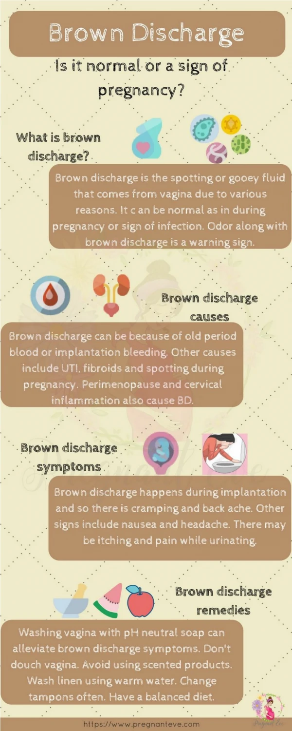
PRESENTING COMPLAINT • Continuous vaginal bleeding 3 month • Foul smelling vaginal discharge 3month
Gynecological history • Menarche 14 years • Past menstrual cycle Regular 6/28 • LMP 21-6-15 • Dysmenorrhea • IMB
Systemic inquiry Not significant
Past Medical and Surgical History Not significant • Family History ovarian and endometrial CA in paternal aunts. hypertension and diabetes • Personal and Socioeconomic history Low socioeconomic group.
General physical examination Pulse 84 /min BP 120/70 Temp 98.6°F R/R 16/min Pallor + Lymph nodes Not Palpable
Systemic examination • ABDOMINAL EXAMINATION • Soft, non-tender • No palpable mass • No visceromegaly • BS audible • CARDIOVASCULAR SYSTEM S1+S2+0 • RESPIRATORY SYSTEM Bilateral vesicular breathing with no added sound • CENTRAL NERVOUS SYSTEM Intact
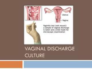
Local examination • Copious amount of foul smelling blood mixed discharge from vagina. • Pelvic examination not done • Rectal examination not done
INVESTIGATIONS • BLOOD CP: Hb 9.9 (12-16 g/dL) TLC 9300 (4000-11000/µL) Plt 323000(150000-450000/µL) • CRP: 3.0 (<6.0mg/l) • Serum sodium: 135 (135-145mmol/L) • Serum potassium: 3.7 (3.5-5.0mmol/L) • HBsAg: negative • Anti HCV: negative
INVESTIGATIONS • LIVER FUNCTION TESTS: Total Bilirubin 0.6mg/dL (upto 1.0) ALT 27 U/L (upto 43) Alk Phos 300U/L (65-360) • RENAL FUNCTION TESTS: Urea 27mg/dL (12-45) Creatinine 0.6mg/dL (upto1.2) Uric acid 3.1mg/dL (2-6)
Usg Private A hypoechoic mass in left adnexa or vagina of 8.5x5.6x 7.1cm Radiology department • Uterus size was 5×3×2cm. • Vagina was dilated, occupied by large hypoechoic, solid mass of 8.5×6.0×6.5cm. • Both adnexae were normal
Tumour markers • B-HCG < 1.20miu/ml (<5miu/ml) • AFP 1.09ng/ml (upto 8.4ng/ml)
Differential diagnosis • Cervical polyp. • Cervical fibroid. • Vaginal growth. • Foreign body. • Hematocolpus.
management • Admission • Plan • EUA and biopsy • Baseline investigations. • One unit of RCC was transfused. • CT scan of abdomen & pelvis
Eua and biopsy • Growth filling vagina • Reddish blue • Necrotic , Friable • Foul-smelling • Arising from cervix • Not attached to vagina • Bleed to touch • Specimen sent to AFIP and DHQ hospital
Post operative • I/V antibiotics were given for 5 days. • Vaginal packing was removed after 24 hours. • Perineal washdown done for 5 days.
Ct scan abdomen & pelvis • Post operative (after 1 week) FINDINGS: • Mild fluid collection • Peri lesional soft tissue stranding . • Fluid was noted in the endometrial canal. • Rest of the findings were normal.
MACROSCOPIC APPEARANCE: Multiple dark brown pieces of tissue collectively measuring 10x5x2 cm.
histopathology MICROSCOPIC DESCRIPTION: The section reveal sheets of small round cells having small oval nuclie and scanty cytoplasm in a hemorrhagic background. Mitosis : 8/10 HPF Necrosis: 20% IMMUNOHISTOCHEMISTRY: Desmin: Positive Ki-67 : 70-80%
Diagnosis EMBRYONAL RHABDOMYOSARCOMA
Referral • NORI hospital for further management. • Slides were sent to POLYCLINIC pathology department and SHAUKAT KHANAM for re-confirmation.
FOLLOW UP ct scan After 3 months Findings • Collection of mixed fluid and blood density in the vagina measuring 9.6×7.9×7.9 cm. • Both ovaries were normal
Mri pelvis • A large well defined mass lesion in the pelvic cavity Centered upon the lower part of uterus and cervix • Extending up to the lower abdomen. • Anteriorly, abutting the posterior wall of the urinary bladder • Posteriorly, compressing the rectum. • Fat planes were not properly visualized.
chemotherapy Three cycles of chemotherapy.
Ct scan(AFTER 3 CYCLES) • A large heterogeneous mass in pelvic cavity measuring 8.5x6.5x6.3 cm • Centered upon the lower part of uterus and cervix • Extending upto lower abdomen till S1.
Ct scan • Anteriorly abutting urinary bladder • Posteriorly pressing on rectum • Fat planes indistinct • No lymphadenopathy • Bony tissue and soft tissues intact
Chemotherapy • Another 3 cycles were given
USG PELVIS USG PELVIS (1 WEEK AGO) • Solid cum cystic mass of 4.7x4.9cm in continuity with cervix. • Well defined and centered in mid pelvis most likely uterine mass
Rhabdomyosarcoma By Dr.Attia Rabbani Senior Registrar Obs/Gynae DHQ Hospital
introduction “A highly malignant tumor of striated muscles derived from primitive mesenchymal cells”