VAGINAL DISCHARGE + PRURITIS
510 likes | 1.75k Views
VAGINAL DISCHARGE + PRURITIS. By: Dr. Samaa Nazer Assistant Professor & Consultant Dept. of Obs/Gyne. Vaginal Discharge. Vaginal discharge may be blood stained white cream, yellow, or greenish discharge and wrongly called leukorrhea.

VAGINAL DISCHARGE + PRURITIS
E N D
Presentation Transcript
VAGINAL DISCHARGE + PRURITIS By: Dr. Samaa Nazer Assistant Professor & Consultant Dept. of Obs/Gyne
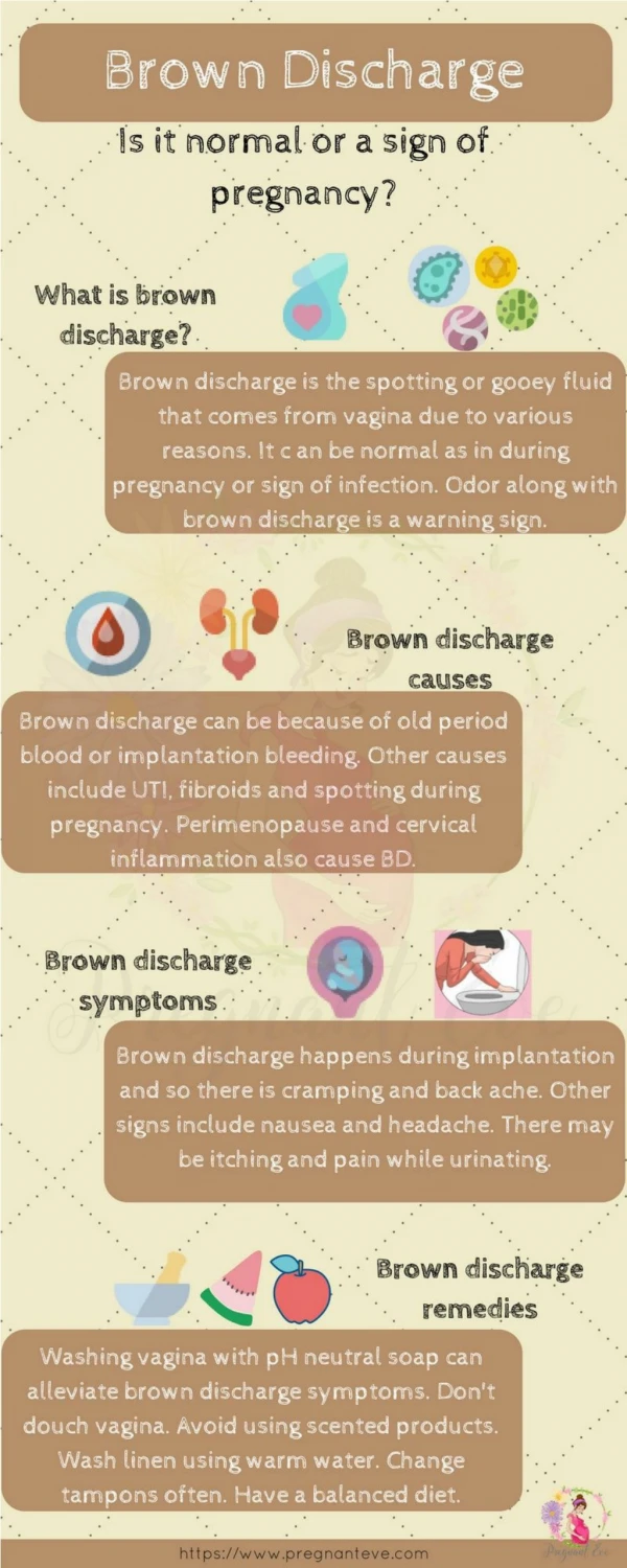
Vaginal Discharge • Vaginal discharge may be blood stained white cream, yellow, or greenish discharge and wrongly called leukorrhea. • Leukorrhea: Excessive amount of normal discharge, never cause pruritus or bad odor. The color is white.
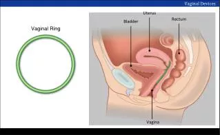
PHYSIOLOGY OF THE VAGINA • The vagina is lined by non-keratinized stratified squamous epithelial influenced by estrogen and progesterone • In children the pH of the vagina is 6-8 predominant flora is gram positive cocci and bacilli • At puberty, the vagina estrogenized and glycogen content increase.
Lactobacilli (Duoderline Bacilli) Convert glycogen to lactic acid pH of the vagina is 3.5-4.5
Vaginal Ecosystem • Dynamic equilibrium between microflora and metabollic by products of the microflora, host estrogen and vaginal pH • The predominant organism is aerobic
Factors affecting the vaginal Ecosystem • Antibiotics • Hormones or lack of hormones • Contraceptive preparations • Douches • Vaginal Medication • Sexual trauma • Stress • Diabetes Mellitus • Decrease host immunity – HIV + STEROIDS
Vaginal Desquamated Tissue • Reproductive age – superfacial cells (est) • Luteal phase- Intermediate cells (prog) • Postmenopausal women- parabasal cells ( absence of hormone)
Differential Diagnosis • Pediatrics + Peripubertal • Physiological leukorrhea – high estrogen • Eczema • Psoriasis • Pinworm- rectum itchy • Foreign body
Investigation: • Swab for culture • PR Examination • EUA • X-RAY pelvic • Exclude sexual abuse • Management: • Hygiene • Antibiotics • Steroids
Post Menopausal • Exclude malignancy
3. Reproductive Age: 1. Physiological : • Increased in pregnancy and mid cycle. • Consists of cervical mucous endometrial and oviduct fluid, exudates from Bartholin’s and Skene’s glands exudate from vaginal epithelium.
2. Infection: • Trichomonas vaginalis • Candida vaginitis • Bacterial vaginosis( non specific vaginitis) • Sexual transmitted disease • Neisseria gonorrhea, chlamydia trachomatis, acquired immune deficiency syndrome, syphilis
3. Urinary and faeculent discharge – vvv 4. Foreign body: IUCD, neglected pessay, vaginal diaphragm 5. Pregnancy: PRM 6. Post cervical cauterization
DIAGNOSIS • History: • Age • Type of discharge • Amount • Onset (relation to antibiotics medication relation to menstruation) • Use of toilet preparation • Colour of discharge • Smell • Pruritus ASSOCIATED SYMPTOMS
2. General Examination:(Anemia, Cachaxia) • Inspection of vulva • Speculum examination • Amount, consistency, characteristic, odor • Bimanual examination
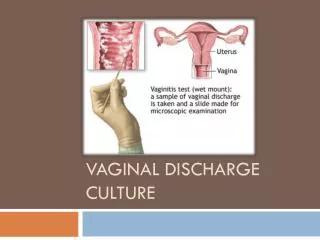
Investigation • 3 Specimens a. Wet mount smear (ad saline)b. Swab for culture and sensitivity c. Gram stain 2. Biopsy from suspicious area 3.Serological test 4. Test for gonorrhea 5. Cervical Smear 6. X-ray in children
Treatment: According to the Cause • Foreign body – remove • Leukorrhoea a. Reassurance b. Hygience c. Minimize pelvic congestion by exercise
Vaginal Infection • Trichomonas vaginitis: • STD: 70% of males contract the disease after single exposure Symptoms: • 25% : asymptomatic • Vaginal discharge , profuse , purulent, malodorous, frequency of urine, dysparunea, vulvar pruritis
Signs: • Thin • Frothy • Pale • Green or gray discharge • pH 5-6.5 • The organism ferment carbohydrates – Produce gas with rancid odor • Erythcum, edema of the vulva and vagina , petcchiea or strawberry patches on the vaginal mucosa and the cervix
Investigation • Identify the organism in wet mount smear • The organism is pear-shaped and motile with a flagellum • Cervical smear • Culture • Immuno-fluorescent staining
Management • Oral Metronidazole (flagyl) • Single dose 2 gm • 500 mg P.O twice for 1 week : • Cure Rate: 95%
Causes of Treatment Failure: • Compliance • Partner as a reservoir Treatment: • Vaginal Route Note: Treatment during pregnancy + Lactation
Candida Vaginitis: Moniliasis • Causative organisms: Candida albicans • Is not STD • CAUSES: • Hormonal factor ( O.C.P) • Depress immunity, diabetes mellitus, debilitating disease • Antibiotics – lactobacilli • Pregnancy estrogen • Premenstrual + Postmenopausal
Symptoms: 20% asymptomatic • Pruritus • Vulvar burning • External dysuria • Dyspareunia • Vaginal discharge ( white, highly viscous, granular, has no odor)
Signs • Erythema • Oedema • Excoriation • Pustules • Speculum: cottage cheese type of discharge • Adherent thrush patches attached to the vaginal wall - pH is < 4.5
Investigation 1.Clinical 2. pH of the vagina norma < 4.5 3. Fungal element either budding yeast form or mycelia under the microscope 4. Whiff test is negative 5. Culture with Nickerson or Sabouraud media (Candida tropicalis)
Management • Standard • Topically applied azole ( nystatin) - 80% - 90% relief 3. Oral antifungal (Fluconazole) 4. Adjunctive treatment topical steroid - 1% hydrochortisone
RECURRENT DISEASE • Definition: More than 3 episodes of infection in one year. • Causes: • Poor compliance • Exclude diabetes mellitus • Candida tropicalis –Trichomonas glabrata
Treatment • Clotrimazol single supp. 500 mg Postmenstrual for 6 months • Oral antifungal: Daily until symptoms disapppear • Culture discharge for resistant type
BACTERIAL VAGINOSIS • STD: • Causative organism: Past Haemophilus or Corynebacterium vaginale • Now: Gardnella vaginalis Gram Negative Bacilli
SIGNS AND SYMPTOMS Symptoms: • 30-40% asymptomatic • Unpleasant vaginal odour (musty or fishy odor) • Vaginal discharge: thin, grayish, or white Signs: • Discharge is not adherent to the vagina, itching, burning is not usual
Diagnosis: • pH: 5-6.5 • Positive odor test- mix discharge with 10% KOH – fishy odor(metabollic by product of anaerobic amins the Whiff test) • Absence of irritation of the vagina and vulvar epithelium • Wet smear – clue cells -Vaginal epithelial cells with clusters of bacteria adherent to their external surface (2% - 5%). -Wet smear shows absent and lack of inflammatory cells.
Complication • Increase risk of pelvic inflammatory disease • Post operative cuff infection after hysterectomy • In pregnancy, it increase the risk of premature rupture of membrane • Premature labour, chorioamnionitis, endometritis
Management • Metronidazole 500 mg twice daily for 7 days Cure is 85% it fall to 50% if the partner is not treated • Clindamycine 300 mg twice daily • Vaginal
Recurrent Causes: • Causes: • Partner • STD • Treatment During Pregnancy:?? The organism may predispose to PRM
PRURITUS VULVAE • Definition: • Means sensation of itching. It is a term used to describe a sensation of irritation from which the patient attempts to gain relief by scratching. • Vulvar irritation: Pain, burn, tender
CAUSES: • Pruritus: associated with vaginal discharge e.g. candida and trichomonas vaginalis. Other discharge which is purulent and mucopurulent discharge cause pain. • Generalized pruritis: Jaundice, ureamia, drug induced • Skin disease specific to vulva: Psoriasis, seborrhoed dermatitis, scabies, Paget’s disease, squamous cell carcinoma • Disease of the anus and rectum: Faecal incontinence, tread worms
Urinary condition: Incontinence: glycosuria • Allergy and drug sensitivity : soaps, deodorant, antiseptic contains phenol, nylon underwear • Deficiency state, Vitamin A, B, B12 , hypochromic macrocytic anaemia • Psychological factor • Chronic vulvar dystrophies : Leukoplakia, lichen sclerosus, Kyourosis vulvae and primary atrophy senile atrohy
1. Investigation 1. History • The onset, site, duration • Presence or absence of vaginal discharge • History of allergic disorders • Medical disease,family history of D.
2. Examination • General – anemia, jaundice • Local examination • Urine for sugar and bile • Blood sugar and liver function test • Bacteriological examination of vaginal discharge • Biopsy from any abnormal vulvar lesion
Treatment • General measure: • Wearing loose fitting • Cotton under clothes • Keep vulva dry and clean regularly • Systemic antihistamine • Local fungicides • Hydrocortisone and local hydrocorticosteroid • Oral antifungal (perianal pruritis) • Estrogen cream • Surgical measure: Local anesthetics, injection, denervation of the vulva , simple vulvectomy