Download

1 / 26
300 likes | 875 Views
Muscles of Thigh. Dr. Sama ul Haque. Objectives. Know the type and formation of hip joint. Differentiate the stability and mobility between the hip joint and shoulder joint. Identify the muscles that act at the hip joint.

E N D
Muscles of Thigh Dr. Sama ul Haque
Objectives • Know the type and formation of hip joint. • Differentiate the stability and mobility between the hip joint and shoulder joint. • Identify the muscles that act at the hip joint. • Identify the muscles of the thigh in terms of their origin, insertion, nerve supply and actions. • Explain the relationships of contents of the femoral triangle to each other & to the surrounding bone and soft tissue landmarks.
Hip Joint Lateral View. Shoulder Lunate surface articulates with head of femur.
Hip joint: articular capsule Anterior Posterior ischiofemoral iliofemoral crest line pubofemoral Note: neck is bare here • Fibrous capsule: • Pubofemoral (medial), resists over abduction • Iliofemoral(anterior), resists hyperextension • Ischiofemoral(posterior), resists hyperextension
Hip joint: articular capsule Blood supply to femoral head: -Retinacular arteries (from medial and lateral circumflex femoral arteries, branches of profunda femoral artery). -Artery of ligament of head (acetabular branch of obturator artery) [deeper orbicular fibres of fibrous capsule] [Synovial membrane: reflects onto neck of femur] Orbicular Fibres Retinacular Arteries artery of ligament of head
Thigh • Three Compartments: Anterior, Medial and posterior • Lateral thigh consists of thickened fascia of the lower extremity called the Fascia Latae or Iliotibial Tract that serves as an insertion of the Tensor Fascia Latae muscle.
Thigh • Anterior compartment: knee extensors and some hip flexors; innervated by femoral nerve, blood supply by femoral artery and its branches. • Medial Compartment: Hip adductors (some rotation and flexion); innervated by obturator nerve and its branch, blood supply by branches of deep femoral artery and obturator artery. • Posterior compartment: Hip extensors and knee flexors; innervated by tibial or common peroneal nerves, blood supply by deep femoral artery.
Functional compartments of the thigh Hip Flexion Knee-extension Hip Adduction Hip Extension Knee-flexion
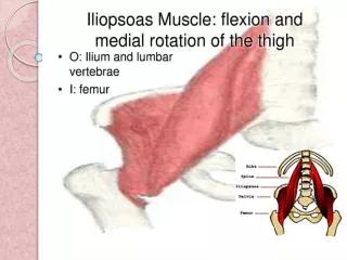
Hip Flexion • Chief flexor of HIP: • Iliopsoas • Psoas major • iliacus • Insertion – lesser trochanter • Femoral nerve (L2-L4): • - Main innervation of • anterior thigh. • Obturator nerve (L2-L4): • - Main innervation of • medial thigh.
pectinius vastus intermedius • ANTERIOR THIGH. • Flexors of hip / Extensors of knee. • Sartorius • Tensor fascia lata • Pectinius • Rectus femoris • Vastusmedialis • Vastusintermedius • Vastuslateralis • Quadriceps femoris = • rectus femoris + vasti • inserts into tibia via • patella • (patellar ligament) (tibial tuberosity) tensor fascia lata sartorius rectus femoris vastuslateralis vastus medialis
Hip extension Gluteus maximus Tensor Fasciae Latae iliotibial tract Gluteal region: -Gluteus maximus (most powerful extensor, also lateral rotator) Insertion: Gluteal tuberosity + Iliotibial tract (band) gluteus maximus FYI Gluteus Maximus and Tensor Fascia Lata insert into Iliotibial Tract - Iliotibial tract is a thickening of the deep fascia (fascia lata) that extends from the ilium to the tibia. - Tension from contraction of gluteus maximus and tensor fasciae latae stabilizes the lower limb as a weight-bearing column.
Posterior Compartment • “Hamstrings” • Common Origin • Medial and Lateral Insertions
Posterior Compartment of thigh: Hamstring muscles. -Extend hip -Flex knee -Common origin at ischial tuborosity. -Innervated by sciatic nerve sciatic nerve semimembranosus semitendinosus biceps femoris Posterior fibres of adductor magnus: Origin from ischialtuborosity, supplied by sciatic nerve, extend hip.
Common origin of extensors Hamstrings. Two insert on medial side: - semimembranosus - semitendinosus (Tibia) Two insert on lateral side: - biceps femoris (Fibula)
Medial Compartment • Muscles • Gracilis, Adductor Longus, Adductor Brevis, Adductor Magnus • Common actions • Pulled groin
Hip Adduction • Medial Compartment • main function = adduction • Obturator externus • Adductor brevis • Adductor longus • Adductor magnus • Gracilis • Most innervated by: • Obturator nerve (L2-L4) • (lumbar plexus) • Exception: • -Hamstring component of • adductor magnus (extensor) • (tibial division of sciatic nerve) obturator externus obturator nerve adductor brevis Adductor magnus adductor longus gracilis
Lateral Rotation of the hip gluteus medius gluteus maximus gluteus minimus Deep to gluteus maximus: -abductors: gluteus medius gluteus minimus (anterior fibres medially rotate) -lateral (external) rotators: piriformis obturatorinternus (associated gemelli) quadratusfemoris [obturatorexternus is also a lateral rotator] piriformis superior gamellus obturator internus quadratus femoris inferior gamellus
iliopsoas pectinius femoral nerve femoral vein femoral artery sartorius adductor longus Femoral Triangle • Boundaries: • Inguinal ligament • Sartorius (lateral) • Adductor longus (medial) • Floor: • Iliopsoas, pectinius, adductor longus • Contents: • Femoral nerve • Femoral artery & deep (profunda) • femoral branch • Femoral vein • Great saphenous vein (superficial), • draining into femoral vein • Lymphatics
Femoral vessels are enclosed by a fascial sleeve [femoral sheath] which is deep to the deep fascia [fascia lata] Lymphatics are found medial to the femoral vein [femoral canal]
Summary: Movements of the Hip Joint (ball and socket). Flexion - Anterior + medial compartments of thigh (iliopsoas, sartorius, rectus femoris, adductor group) Extension - Gluteal region /posterior compartment of thigh (gluteus maximus, hamstrings, adductor magnus) Adduction - Medial (adductor) compartment of thigh Abduction - gluteus medius & minimus, Tenor Fascia Lata Rotation: Lateral - Gluteus maximus, lateral rotators Medial - anterior parts of gluteus medius & minimus, + Tensor Fascia Lata
Blood Supply • Femoral Artery • Deep Femoral (Femoral Profunda) • Medial Circumflex • Lateral Circumflex • Ascending Branch • Lateral Branch • Descending Branch