Download
1 / 84
870 likes | 1.31k Views
Learn about full blood count, anemia, iron deficiency, differential diagnosis, macrocytic anaemia, myelodysplasia, and more. Includes practical classifications, investigations, and management approaches.

E N D
Haematology in Primary Care:The Full Blood Count Charles Crawley George Follows Cambridge Haematology Partners www.cambridgehaematology.com
The FBC www.cambridgehaematology.com
Haemoglobin • Low haemoglobin defines anaemia • Males 13-18g/l • Females 11.5-16g/l • Variations: • Children • Neonates – 14-24g/l • 2 months – 8.9-13.2g/l • 9-12ys - 11.5-15.4g/l • Pregnancy • 3rd Trimester – 9.8-13.7g/l • Age • 5-7th decade – falls in men rises in women • Exercise • Increases Hb • Altitude • Smoking www.cambridgehaematology.com
MCV • Mean Cell Volume: average size of RBC • Normal adult : 76 (80) - 100 fL • MCV < 76 fL (microcytic) • MCV > 100 fL (macrocytic) • MCV 80 - 100fL (normocytic) www.cambridgehaematology.com
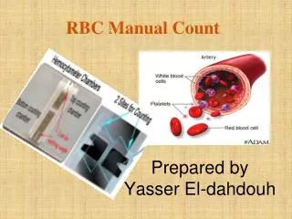
Practical Classification of Anaemia • Reticulocyte count: In the investigation of anaemia • Reduced: Failure of erythropoiesis • Increased: Appropriate BM erythroid response www.cambridgehaematology.com
35 year Male • Hx: Lethargy, SOB • Sx: Pale • FBC: Hb 6.4 g/dL MCV 71 fL RDW 0.19 WCC 5.2 Platelets 375 • Film: Severe hypochromasia and microcytosis www.cambridgehaematology.com
Commonest Causes Iron Deficiency www.cambridgehaematology.com
Blood Film www.cambridgehaematology.com
Differential Diagnosis • Causes of microcytic hypochromic anaemia • Iron deficiency • Blood loss • Malabsorption - Coeliac disease; gastrectomy • Increased utilisation - parasites • Dietary deficiency - rare • Haemoglobinopathy • Anaemia of chronic disease www.cambridgehaematology.com
Fe Deficiency vs Anaemia of Chronic Disease GIT studies: endoscopy etc www.cambridgehaematology.com
26 year Female • Hx: Antenatal visit; First trimester • FBC: Hb 11.0 g/dL MCV 73 fL MCH 27 pg RDW 0.14 WCC 8.5 x 10⁹/L Platelets 164 x 109/L • Film: Microcytic RBC www.cambridgehaematology.com
Fe Deficiency vs Hbinopathy Check iron status: Ferritin Family history / ethnicity: Thalassaemia / haemoglobinopathy Need to determine risk to fetus of severe thalassaemic syndrome (in 1st rimester): Homozygous thalassaemia (α or β) Homozygous Hb S (sickle cell disease) Severe compound heterozygous states E.g.: HbS/β; HbE/β; HbSC Determine need to check partner www.cambridgehaematology.com
Red Cell Distribution Width (RDW) • The degree of variation in size of RBC: N <14 • Increased RDW corresponds with anisocytosis: • Iron deficiency (increased RDW is the earliest lab feature: anisocytosis precedes the anaemia) • Megaloblastic anaemia (can be very high >20) • Anaemia with bone marrow erythroid response (i.e. reticulocytosis) • RDW useful in DDx of microcytic anaemias. • Most cases of iron deficiency: raised RDW • Most cases thalassaemia trait: normal RDW www.cambridgehaematology.com
MCH • Mean Cell Haemoglobin (27-32 pg) • The mean haemoglobin per red blood cell • MCH usually rises or falls as the MCV is increased or decreased. • MCH < 25 pg used as a guide to the presence of thalassaemia or haemoglobinopathy. • MCH usually markedly reduced in thalassaemia (e.g. beta thalassaemia trait MCH 19 pg) www.cambridgehaematology.com
Haemoglobin Studies 1. Normal adult 2. HPFH (heterozygote)3. Hb S--HPFH 4. Hb C--HPFH 5. Normal newborn A/F/S/C control www.cambridgehaematology.com
73 year male • Hx: Tiredness • FBC: Hb 4.0 g/dL MCV 102 fL RDW 0.24 WCC / Plt Normal • Film: Macrocytes, fragmented red cells, occasional NRBC www.cambridgehaematology.com
Blood Film73 yr old male www.cambridgehaematology.com
Severe Macrocytic Anaemia • Megaloblastic anaemia • Liver disease: end-stage failure • Red cell aplasia: • Parvovirus; thymoma, other malignancy • Bone marrow failure or infiltration: • Myelodysplasi • Multiple myeloma www.cambridgehaematology.com
Investigations 1. Serum vitamin B12 Red cell folate (serum folate) 2. Reticulocyte count (BM erythroid function) 3. Liver function 4. Parvovirus serology 5. Bone marrow examination www.cambridgehaematology.com
Don’t Forget the Alcohol www.cambridgehaematology.com
Other Causes of Macrocytic Anaemia • Severe liver disease • Excess alcohol • Haemorrhage / haemolysis: reticulocytosis • Drug therapy: esp. cytotoxics • Hypothyroidism • Myelodysplasia • Marrow infiltration • Bone marrow examination may be indicated www.cambridgehaematology.com
Myelodysplasia (MDS) • Clonal disorder • Ineffective haematopoiesis • Incidence increases with age • Age 50yrs - 1 per 100,000 • Age 70yrs – 25 per 100,000 • RBC, WCC, and platelets affected www.cambridgehaematology.com
Myelodysplasia Bone Marrow Peripheral Blood www.cambridgehaematology.com
Myelodysplasia • Prognosis • Number of cytopenias • BM Blast percentage • Cytogenetics • Age • Survival • Varies 11.7 yrs – 0.4yrs • Management • Supportive • Stem cell transplantation • New drugs www.cambridgehaematology.com
Normocytic Anaemia • Multiple aetiologies • Primary marrow production defect • Myelodysplasia • Marrow infiltration • Haematinic deficiencies • Reduced red cell survival • Blood loss • Intrinsic defects (eg. Enzyme; membrane) • Extrinsic defects (eg. Plasma problems) www.cambridgehaematology.com
Approach to Normocytic Anaemia • History: • Acute blood loss ; jaundice; dark urine • Exclude treatable causes: • Check ferritin, folate, vitamin B12 • Renal and hepatic function • Acute phase reactants • Consider haemolysis The blood film may have the answer ! www.cambridgehaematology.com
78 year male • Hx: Chest pain • PMHx: Myocardial infarct • FBC: Hb 7.2 g/dL MCV 97 fL WCC 4.5 x 10⁹/L Platelets 320 x 109/L Reticulocytes: 320 (10-100) www.cambridgehaematology.com
Blood Film www.cambridgehaematology.com
Blood Film • RBC: Spherocytes Polychromasia Nucleated red cells Spherocytic haemolytic anaemia: Auto-immune haemolytic anaemia Hereditary spherocytosis www.cambridgehaematology.com
Other Investigations • Biochemistry: • Bilirubin 100 μmol/L (<20) • Other LFT Normal • LDH 1,500 U/L (120-240) • Haptoglobin <0.1 • Haematology: • Reticulocyte count • Direct anti-globulin (Coombs) test: Positive • Enzymes, • Hereditary spherocytosis screen www.cambridgehaematology.com
Primary Red Cell Problem: Red cell membrane: Hereditary spherocytosis Enzyme defect: G6PD deficiency Haemoglobin defect: thalassaemia Abnormal red cells: dyserythropoiesis (MDS) Secondary Red Cell Destruction Autoimmune Severe hepatic dysfunction Red cell fragmentation: DIC; HUS; TTP Infections: malaria; clostridium Haemolytic Anaemia www.cambridgehaematology.com
Blood Film blister or helmet cells Glucose-6-phosphate dehydrogenase deficiency www.cambridgehaematology.com
Normocytic Anaemia • Blood film may have the answer: • Normal red cell morphology • Dimorphic (high RDW): 2x RBC populations • Marked anisocytosis: marrow dysfunction/MDS • Is there polychromasia? • Yes: Anaemia with marrow response • No: Impaired marrow response • Anaemia of Chronic Disease • BM failure • Red cell aplasia: Parvovirus; aplastic anaemia www.cambridgehaematology.com
Polycythaemia • Pseudopolycythaemia • Primary • Polycythemia vera • Secondary • Hypoxia • Altitude • Cardiac/Pulmonary disease • Cirrhosis • Abnormal Haemoglobins • Chronic CO exposure • Inappropriate erythropoietin • Renal lesions • Tumours • Drug www.cambridgehaematology.com
Clinical Features • Hyperviscosity • Headaches • Blurred vision • Breathlessness • Confusion • (Plethora) • Thrombosis • Venous + arterial • Bleeding • Other • Pruritis • Gout www.cambridgehaematology.com
Polycythaemia investigations • FBC + Film • CXR • Cardiac assessment • Red Cell mass • Blood gasses • Major advance – JAK 2 mutation screens www.cambridgehaematology.com
JAK2 • Presence of the V617F mutation indicates that the patient has an acquired, clonal hematological disorder and not a reactive or secondary process. • Absence of the JAK2 V617F mutation does not exclude a MPD as up to 50% of patients with ET and IMF will have wildtype JAK2. • The V617F mutation does not help in sub-classifying the type of MPD of a given patient www.cambridgehaematology.com
Pathogenesis: Deregulated Tyrosine Kinases in MPD CML BCR-ABL CMML TEL-PDGFRB CEL FIP1L1-PDGFRA SM KIT D816V PV JAK2 V617F ET JAK2 V617F IMF JAK2 V617F www.cambridgehaematology.com
Questions so far? www.cambridgehaematology.com
Platelets • Too many (thrombocytosis) • Too few (thrombocytopenia) • Dysfunctional • When should we worry? www.cambridgehaematology.com
Thrombocytosis • > 450 x 109/l • Causes • Reactive (almost anything!) • Common – bleeding, infection, malignancy • Tend to be less than 1000 x 109/l • Primary bone marrow disorder • Myeloproliferative disorders (up to 3000+) (essential thrombocythaemia, myelofibrosis, polycythaemia, chronic myeloid leukaemia) www.cambridgehaematology.com
Thrombocytosis • History, examination should guide investigations and referrals • Should the patient be on aspirin? • Only firm evidence is MPDs • Reactive often given if > 1000, but little evidence for this www.cambridgehaematology.com
Thrombocytosis • Essential Thrombocythaemia (ET) • Long term management balancing thrombotic vs bleeding risk • Aspirin for intermediate risk • Cytoreduction + aspirin for higher risk (beware the pseudohyperkalaemia!!) www.cambridgehaematology.com
Thrombocytopenia • Is it real??? • Poor sample • Clumped in EDTA – blood film **If at all possible confirm with a repeat sample www.cambridgehaematology.com
Thrombocytopenia Pancytopenia vs Isolated low platelets • Pancytopenia – • always serious (marrow failure) • Isolated • may be relatively unimportant www.cambridgehaematology.com
Thrombocytopenia • Decreased production • Rare in isolation • Viral infections • Increased consumption • Autoimmune – ITP • Drugs • Pregnancy • Large spleen / portal hypertension • Infections (HIV) • RARE but serious TTP – HUS - DIC www.cambridgehaematology.com
Thrombocytopenia • Investigations depend on clinical suspicion • Platelet volume may be helpful (small - think marrow) • <8.0 and >10.5 • BM often not required www.cambridgehaematology.com
Thrombocytopenia (not joint bleeds!) www.cambridgehaematology.com
Thrombocytopenia • How real is the bleeding risk? • Cause of low platelets • Wet vs dry purpura • Platelet transfusions often not useful www.cambridgehaematology.com
ITP – a few useful reminders • Children • Acute, post viral, • spontaneous resolution (often no therapy) • Adult • More insidious onset • Chronic = common • Many (not all!) cases do require treatment • Steroids – splenectomy • Novel therapies www.cambridgehaematology.com