Pulse Oximetry
Pulse Oximetry. Theodore Maniatis MD Director of Critical Care. This module was developed by Theodore Maniatis MD, FACP, FACCP, the Director Critical Care and is intended as an educational resource for all Registered Professional Nurses. Co-Oximetry.

Pulse Oximetry
E N D
Presentation Transcript
Pulse Oximetry Theodore Maniatis MD Director of Critical Care
This module was developed by Theodore Maniatis MD, FACP, FACCP, the Director Critical Care and is intended as an educational resource for all Registered Professional Nurses.
Co-Oximetry To understand pulse-oximetry one should first understand about co-oximetry.
Co-Oximetry • Oxyhemoglobin • Deoxyhemoglobin • Carboxyhemoglobin • Methemoglobin Co-oximetry is the ability to measure the different types of hemoglobin in an arterial blood sample including oxygenated hemoglobin known as oxyhemoglobin, deoxygenated hemoglobin or deoxyhemoglobin, carboxyhemoglobin, and/or methemoglobin. It is an invasive way of measuring oxygen saturation.
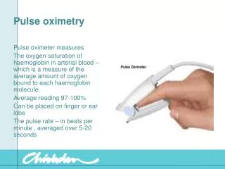
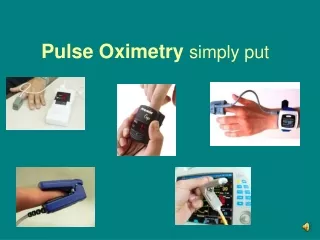
Pulse Oximetry Pulse Oximetry is a non-invasive method to monitor the percentage of hemoglobin that is saturated with oxygen.
Oxygen Transport • Oxygen is carried in blood • Released to tissues by diffusion • It moves from an area of high partial pressure →low partial pressure • Tissues continuously use oxygen for energy • O2 concentration in tissues is always low stimulating hemoglobin to release oxygen to the tissues
Oxyhemoglobin 98.5% of Oxygen is carried bound to hemoglobin It is important to know the quantity of oxyhemoglobin because 98.5% of oxygen is carried bound to hemoglobin as a form called oxyhemoglobin. Only 1.5% of oxygen is dissolved in blood and is described as the PaO2 of the blood. This is the only form that diffuses across cell membranes.
Spectrophotometric Method of Oxygen Measurement In the absence of other materials that absorb light at the same wavelength, the concentration of a substance in solution is proportioned to the amount of light absorbed. • The spectrophotometric method of measuring the different types of hemoglobin is the most commonly used method on today’s modern analyzers. The principle behind the physics of the spectrophotometric method states that in the absence of other materials that absorb light at the same wave length, the concentration of a substance in solution is proportional to the amount of light absorbed. • The spectrophotometric method transmits light through an arterial blood sample and depending on the wave length of the light transmitted through the sample one can measure the different types of hemoglobin since each type of hemoglobin absorbs light of a different wave length.
Light Absorption • Deoxyhemoglobin absorbs light maximally in the red band of the spectrum (600-700 nm) (660 nm) • Oxyhemoglobin absorbs light maximally in the infrared band of the spectrum(850-1000 nm) (940 nm)
Because deoxyhemoglobin absorbs light maximally in the red band of the spectrum at about 660 nanometers and oxyhemoglobin absorbs light maximally in the infrared band of the spectrum at about 940 nanometers one can shine a light through a blood or tissue sample at the 660 nanometer band and at the 940 nanometer band and measure the quantities of oxyhemoglobin and deoxyhemoglobin.
There is both oxygenated and deoxygenated hemoglobin in venous blood, capillary blood, and arterial blood. There is some absorption of light in tissue and bone. The way the pulse-oximeter measures the amount of oxygenated hemoglobin in arterial blood and disregards the amount of blood present in venous blood, capillary blood and tissue is by recognizing the absorption of light at certain times (the pulse time or the phase of pulsation where the arteries carry more blood). It disregards the absorption that is stable and only measures the absorption that is pulsatile.
Accuracy Between 70% and 100% (2%) • The accuracy of pulse-oximetry when compared to co-oximetry is very good with only a 2% error when the saturations are between 70% and 100%. Between 50% and 70% (3%) • There is approximately a 3% error when the saturations are between 50% and 70%. Clinical impressions however suggest saturations below 80% should be checked with ABG. • Most experts and practicing physician would suggest that when saturations are below 80%, one should check an arterial blood gas.
Artifact • These accuracy statements refer to measurements made when there is no artifact present and no other sources of error present; these are the most consistent and reproducible measurements • that current technology lets us make. • There are sources of artifact when using a pulse-oximeter whether it is a forehead pulse-oximeter, an ear pulse-oximeter or a finger probe pulse-oximeter. • The sources of artifact include improper probe placement, motion artifact, ambient light and electromagnetic radiation. • Improper probe placement • Motion artifact • Ambient light • Electromagnetic radiation.
Hypoperfusion • Anemia (Hgb 5g/dl) • Venous congestion • Skin pigmentation • Hypothermia • Vital dyes • Nail polish (red OK) • Acrylic nails • Sources of error other than artifact include hypoperfusion, hypothermia, anemia (the anemia must be severe usually with a hemoglobin concentration less than 5g/dl) and venous congestion (such as that seen in right heart failure). • Other sources include skin pigmentation (such as that seen in very dark skinned African American patients or patients with intense hyperbilirubinemia), nail polish, remembering that red nail polish doesn’t affect pulse-oximetry significantly. • When it comes to nail polish remember the following: if it’s good, it’s good-- meaning you don’t get a falsely high pulse-oximetry reading from nail polish, only possibly falsely low readings. • Acrylic nails effect the accuracy of pulse-oximetry.
Errors CO absorbs approximately the same amount of 660nm light as does oxyhemoglobin. • Other sources of error which deserve specific mention include carboxyhemoglobin because carboxyhemoglobin absorbs approximately the same amount of light at 660 nanometers as does oxyhemoglobin.
As the true carboxyhemoglobin concentration increases on the x-axis & the amount of oxyhemoglobin, (the blue squares) as measured by co-oximetry is falling, the hemoglobin saturation measured by pulse-oximetry (the red squares) is remaining the same thereby giving a falsely high impression of how much oxyhemoglobin exists.
SIUH’s practice requires co-oximetry on a patient’s first blood gas. • Calculated saturations don’t help. • The way one deals with suspected carbon monoxide exposure is to obtain a co-oximetry test to help avoid misdiagnoses and safeguard patients. • The Staten Island University Hospital practice requires that a co-oximetry be performed on a patient’s first blood gas. • The reason that this is important is because calculated saturations do not help identify the problem. Co-Oximetry
Pulse rate on the oximeter should correspond with patient’s actual pulse. The quality of the signal is represented by the *** on the monitor screen. 3 *** indicate the best signal. Position finger probes so that the light beams and sensors are opposite each other. Disposable probes should not be used on earlobes. Pulse waveform should be displayed. 5 Implications for Nursing
The most common misuse of pulse-oximetry is the result of placing too much reliance on the measurements. For example, this X-ray represents the X-ray of a 61 year old male who presented to the Emergency Department with fever and sharp chest pain for 2 days associated with a cough and shortness of breath. The history in the chart reads that his pulse ox saturation was 97% which sounds OK except that no one noted how much supplemental oxygen, if any, the patient was receiving. If he was receiving no supplemental oxygen this would be a fairly good saturation. If he was receiving 50% oxygen the saturation would be OK but the gas exchange problem would be significant.
This next X-ray is the same patient who has more shortness of breath, yellow sputum production and now has a 91% saturation on room air and 98% saturation on 2 liters. Obviously the 98% saturation is “acceptable” but we have to give supplemental oxygen to the patient today in order to achieve it where as yesterday we did not have to use supplemental oxygen.
This next X-ray represents a patient with a pulse-oximetry of 90% on room air and only 91% on 3 liters, obviously indicating that this patient is having a greater problem achieving saturation above 90% then he was the day before. At this point one can not tell if the patient’s oxygenation problem is associated with hypoventilation which is the rentention of carbon dioxide or hyperventilation with a reduction in carbon dioxide. An arterial blood gas is indicated in this situation to measure the PaCO2 and the Ph. It must be remembered that pulse-oximetry measures one and only one thing; the amount of oxygenated hemoglobin. At this point in time to measure the PaCO2, and the Ph one needs an arterial blood gas. One must also always remember to ask themselves how much oxygen was required to achieve a normal or acceptable oxyhemoglobin level. The more oxygen that is required to keep the oxygen saturation above 90% the worse the patient’s gas exchange problem.
Post-program Evaluation Survey In order to get credit for the completion of this program you are required to do the following: • Click on the link below. This will bring you to the attestation form where you will indicate “Yes, I have read the information in this learning module.” CLICK HERE
Acknowledgements Content and Presentation: • Theodore Maniatis MD, FACP, FACCP Slide design and layout: • Theresa Bucco MSN, RN-BC, PhD-C • Barbara Pfaff MSN, RN, ACNP-BC, CCRN Narration: • Barbara Pfaff MSN, RN, ACNP-BC, CCRN Webpage layout and links: • Teresa Heithaus MSN, RN