Download

1 / 15
220 likes | 774 Views
Basic Patient Monitoring For Anesthesia Pulse Oximetry. Dr. Jeffrey Elliot Field HBSc , D.D.S. , Diplomat of the National dental Board of Anesthesia, Fellow of The American Dental Society of Anesthesia. The Machines That Go Beep. Pulse Oximetry. How Does It work.

E N D
Basic Patient Monitoring For AnesthesiaPulse Oximetry Dr. Jeffrey Elliot Field HBSc, D.D.S. , Diplomat of the National dental Board of Anesthesia, Fellow of The American Dental Society of Anesthesia
How Does It work • The pulse oximeter produces both a red light at 660 nm ( which we can see) and infrared light at 910 nm (which we cant see). • These shine through a thin piece of tissue and the pulse oximeter measures the relative absorption of the two wavelengths. From this it calculates the oxygen saturation, i.e. the ratio of oxyhaemoglobin to deoxyhaemoglobin, from absorption curves programmed into the device.
How Does It work • What makes the pulse oximeter so useful is that it only looks at the pulsatile component of the light absorption, not the background level. The pulsatile change in tissue absorption is due to blood entering the arterioles and therefore the pulse oximeter measures arterial oxygen saturation (Sa02%).
HOW DOES IT WORK/PHYSIOLOGY • Oxygenated hemoglobin absorbs more infrared light and allows more red light to pass through. Deoxygenated (or reduced) hemoglobin absorbs more red light and allows more infrared light to pass through. • Red light is in the 600-750 nm wavelength light band. Infrared light is in the 850-1000 nm wavelength light band.
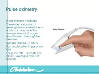
HOW DOES IT WORK. • Pulse oximetry uses a light emitter with red and infrared LEDs that shines through a reasonably translucent site with good blood flow. • Typical adult/pediatric sites are the finger, toe, pinna (top) or lobe of the ear. • Opposite the emitter is a photodetector that receives the light that passes through the measuring site.
WHAT DOES IT TELL US. • The pulse oximeter measures the adequacy of arterial oxygenation. Pulse oximeters are very useful monitors in anesthesia for two reasons. • Firstly, arterial hypoxaemia is the final "common pathway" for many problems that occur in anesthesia such as hypoventilation, airway obstruction and equipment-related problems. A pulse oximeter can therefore pick up problems of different etiologies. • Secondly, hypoxia is a common and serious problem so noticing it early will prevent disasters. A pulse oximeter will detect hypoxemia long before the patient becomes cyanosed ( turns blue).
Normal Values • The normal values for healthy individuals are 95-100% • Please never forget that a reading of 90% correlates with an arterial oxygen saturation of 60%. This value is what we call the edge of the cliff. From 90% on down the patient’s oxygen saturation will drop rapidly. • Also remember that values under 70% ( 40% arterial saturation) are not reliably correlated with arterial oxygen saturation.
Why the Curve Shifts • When the pH is decreased (increased acidity), the affinity of hemoglobin to oxygen is decreased and thus unloading is easier. This is called the Bohr effect. Thus at the tissue level where the pH is low (high level of carbon dioxide), cellular oxygenation is facilitated. As for the oxyhemoglobin dissociation curve, this is shifted towards the right. • The same effect can be seen by an increase in temperature. Again this temperature effect helps in the unloading of oxygen at the tissue level. • red blood cells lack mitochondria, energy is obtained through the anaerobic metabolism of glucose. One metabolite of this reaction is 2,3-diphosphoglyceric acid (2,3-DPG). 2,3-DPG production is increased by an increase in deoxyhemoglobin production. 2,3-DPG, in turn, increases the stability of deoxyhemoglobin and thus shifts the dissociation curve to the right.
Why Is This important? • If the curve is shifted to the right then we can accept lower oxygen saturations as these correlate with higher arterial oxygen saturation. • This issue should not be relevant to the sedationist except in an emergency let us say a resuscitation following cardiac arrest where the patient may have some acidosis or in a case of malignant hyperthermia where you have increased body temperature.
Practical considerations for Pulse oximetry • You must put the probe on in the correct orientation. • That is the part with the shining red light sits over the nail bed not the dorsal aspect of the finger. • Pulse Oximeters measure pulsatile blood flow. Therefore if blood flow is restricted [i.e. The patient is cold or for some other reason there is not good perfusion to the fingers] you will not get an accurate reading.