Download
1 / 1
10 likes | 164 Views
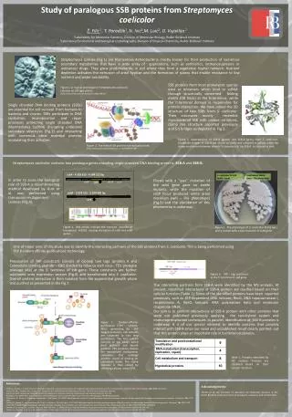
Study of paralogous SSB proteins from Streptomyces coelicolor. Ž. Filić 1 , T. Paradžik 1 , N. Ivić 2 ,M. Luić 2 , D. Vujaklija 1 1 Laboratory for Molecular Genetics, Division of Molecular Biology, Ruđer Bošković Institute

E N D
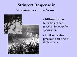
Study of paralogous SSB proteins from Streptomyces coelicolor Ž. Filić1, T. Paradžik1,N. Ivić2,M. Luić2, D. Vujaklija1 1Laboratory for Molecular Genetics, Division of Molecular Biology, Ruđer Bošković Institute 2Laboratory for chemical and biological crystallography, Division of Physical Chemistry, Ruđer Bošković Institute ssbB ssbA - 4 303 613 - 4 304 212 bp rpS6 rpS18 rpL9 ssbA ssbB - 2 924 515 - 2 924 985 bp Streptomyces species (Fig.1)are filamentous Actinobacteria mostly known for their production of numerous secondary metabolites that have a wide array of applications, such as antibiotics, immunosupresors or anticancer drugs. They grow predominantly in soil where they form a vegetative hyphal network. Nutrient depletion activates the extrusion of areal hyphae and the formation of spores that enable resistance to low nutrient and water availability. SSB proteins from most prokaryotic species exist as tetramers which bind to ssDNA through structurally conserved folding motifs (OB folds) at the N-terminus, while the C-terminal domain is responsible for protein interaction.We have solved the 3D structure of two SSBs from S. coelicolor. Their structures mostly resemble mycobacterial SSB with unique variations, clamp like structure reported previously and S-S bridges as depicted in Fig.3. Figure 1. A typical phenotype of Streptomyces coelicolor colonies on SM agar plates. (http://www.flickr.com/photos/ajc1/3288213824/) Single stranded DNA binding proteins (SSBs) are essential for cell survival from humans to bacteria and viruses. SSBsparticipate in DNA replication, recombination and repair processes protecting single stranded DNA intermediates (ssDNA) disrupting undesired secondary structures (Fig.2) and interacting with numerous other essential proteins modulating their activities. Figure 3. Superposition of SSB-B (green) and SSB-A (grey) from S. coelicolor. Disulphide bridges of SSB-B are shown as sticks and coloured in yellow, while the clamp mechanism between strands 9 characteristic for SSB-A is coloured in pink. Figure 2. The role of SSB protein in a replication fork http://www.pdbj.org/eprots/index_en.cgi?PDB%3A3BEP • Streptomyces coelicolor contains two paralogus genes encoding single stranded DNA binding proteins:SSB-A and SSB-B. In order to asses the biological role of SSB-A a recombineering method developed by Gust et al. was performed using transposon-mutagenised cosmids (Fig.4). Clones with a ‘’scar’’ mutation of the ssbA gene gave no viable mutants, while the mutation of ssbB locus produced white areal mycelium (whi – like phenotype) (Fig.5) and the elucidation of this phenomena is underway. S. coelicolor M 145 (wild – type) Inactivated SSB-B (whi – like phenotype) Figure 4. Red arrows indicate the insertion position of transposon tn5062 causing disruption of ssbA and ssbB genes Figure 5. The phenotype of S. coelicolor M145 (wt ) and a mutant with a scar mutation of ssbB gene One of major aims of this study was to identify the interacting partners of the SSB proteins from S. coelicolor. This is being performed using TAP (tandem affinity purification) technology. Preparation of TAP constructs consists of cloning two tags (protein A and Calmodulin binding peptide - CBP, divided by tobacco etch virus - TEV protease cleavage site) at the 5’ terminus of ssb gene. These constructs are further subcloned onto expression vectors (Fig.6) and transformed into S. coelicolor. Overall protein extraction is then isolated from the exponential growth phase and purified as presented in the Fig.7. Figure 6. TAP - tag constructs at the 5’ terminus of ssB gene • The interacting partners from SSB-A were identified by the MS analysis. At present, identified interactants of SSB-A protein are clasified based on their cellular function (Table1). Some of the identified proteins have been reported previously, such as ATP-dependent DNA helicase, RecG, DNA topoisomerase I, recombinase A, RecQ helicase, RNA polymerase beta and molecular chaperone DNaK. • Our aim is to confirm interactions of SSB-A protein with other proteins that were not published previously applying the two-hybrid system and immunoprecipitation techniques. In parallel, identification of SSB-B proteins is underway. It is of our special intrerest to identify proteins that possibly interact with SSB-B since our novel and unpublished result clearly pointed out that this protein plays an important role in bacterial cytokinesis. Figure 7. Tandem-affinity-purification (TAP) scheme. After generating the TAP tagged protein(s), cell extracts are subjected to two step purification. The first column consists of IgG beads which bind proteinA and bound proteins. TEV protease cleaves the immobilized multiprotein complexes that undergo another round of binding on calmodulin beads. The native complex is then eluted by chelating calcium using EGTA. Table 1. Proteins identified by MS analysis. Proteins are classified based on their cellular function. References: Flardh, K., Findlay, K.C. and Chater, K.F. (1999) Association of early sporulation genes with suggested developmental decision points in Streptomyces coelicolor A3(2). Microbiology, 145 ( Pt 9), 2229-2243. Kieser, T.B., M.J.; Buttner, M.J.; Chater, K.F.; . Hopwood, D.A. (2000) Practical Streptomyces Genetics. The John Innes Foundation, Norwich. Gust et al (2004) Lambda red-mediated genetic manipulation of antibiotic-producing Streptomyces. Adv Appl Microbiol, 54, 107-128. Hopwood, D.A. (1999) Forty years of genetics with Streptomyces: from in vivo through in vitro to in silico. Microbiology, 145 ( Pt 9), 2183-2202. Jakimowicz, D., Mouz, S., Zakrzewska-Czerwinska, J. and Chater, K.F. (2006) Developmental control of a parAB promoter leads to formation of sporulation-associated ParB complexes in Streptomyces coelicolor. J Bacteriol, 188, 1710-1720. Lindner, C., Nijland, R., van Hartskamp, M., Bron, S., Hamoen, L.W. and Kuipers, O.P. (2004) Differential expression of two paralogous genes of Bacillus subtilis encoding single-stranded DNA binding protein. J Bacteriol, 186, 1097-1105. Schrempf, H (2008) Streptomycetaceae: Life Style, Genome, Metabolism and Habitats. eLS. John Wiley & Sons Ltd, Chichester. http://www.els.net [doi: 10.1002/9780470015902.a0020393] Shereda, R.D., Kozlov, A.G., Lohman, T.M., Cox, M.M. and Keck, J.L. (2008) SSB as an organizer/mobilizer of genome maintenance complexes. Crit Rev Biochem Mol Biol, 43, 289-318. Stefanic, Z., Vujaklija, D. and Luic, M. (2009) Structure of the single-stranded DNA-binding protein from Streptomyces coelicolor. Acta Crystallogr D Biol Crystallogr, 65, 974-979. Acknowledgements: Thanks to all the members of Laboratory for Molecular Genetics at the Ruđer Bošković institutefor their kind support, assistance and collaboration.