Download
1 / 74
750 likes | 824 Views
Learn about the prevalence, causes, and symptoms of urinary tract infections (UTIs) in children, along with recommended diagnostic methods such as urine culture and bladder cannulation.

E N D
ewa demidowicz Urinary tract infectionsUTI
Urinary tract infections • Definition: Presence of germs in urinary tract above the urinary bladder sphincter
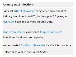
Epidemiology • Urinary system forms the second (after the respiratory system) source of infections regarding their frequency. • Urinary system infections are the cause of about 5% of pyrexic episodes in the case of children <1year of age • UTI occurs in approximately 8% of girls and 2% of boys below 7.
Epidemiology • Below the third of month of life, there is a 5-6 times greater dominance of male sex, which may enventuate from a higher frequency of the occurence of urinary system malformations. • Above the 8th month grows the number of urinary infections among girls • In the case of females, urinary infections develop 10-50 times more frequently than in the case of men.
Etiology • Most frequently Gram-negative bacteria • 80-85% Echerichia coli
Etiology • Apart from that, in cultures are also found: • Proteus mirabilis (most commonly found under the prepuce) • Klebsiella, Enterobacter, Morganella morgani, Pseudomonas aeruginosa • Gram positive bacteria • - Staphylococcus: Staph saprophyticus characterisic for teenage girls, • - Streptococcus spp: UTI mainly in the case of newborns
Epidemiology – other • Fungi – candida albicans, especially after a long-term antibiotic therapy, immunosuppresive treatment, and application of cytotoxic agents • Viruses – adenovirus, causes hemorrhagic cystitis • Chlamydia trachomatis – such etiology should be taken into consideration in the case when aseptic leukocyturii is accompanied by suppurative leakage from the urethra • Trichomonas vaginalis – newborns/infants
98 % of all infections develops in ascending tract . • urethra- bladder – kidney. • UTI is usually result of bowel flora entering the urinary tract via the urethra • 1% of urinary infections develops through blood, together with septicaemia.
symptoms1. Newborns and children <3month Untypical Symptoms • vomiting, diarrhoea, apetite loss, prolonged jaundice, anxiety, polyuria • In severe conditions symptoms of general infection • Only in 40% UTI of children <3month fever is present !
symptoms2. Children from 3 months to 3 years • Fever, apetite loss, nausea ,vomiting, stomach pain, weight gain inhibition, anxiety/pain accompanying urination, change of colour and smell of urine • Rare classical symptoms of dysuria • (bladder tenesmus, frequency, pain and burning while urinating)
Symptoms3. Children >3 years. • Symptoms characteristic for urinary tract infections = dysuria, fever, abdominal pain • Pyelonephritis – fever >38,5degrees, pain in lumbar region, positive Goldflam’s symptom • Cystitis – fever <38,5degrees, dysuria, without the pain in lumbar region or the Goldflam’s symptom • ! In the case of children with recurrence the course is most commonly asymptomatic.
Diagnostics:Laboratory tests1. Urinalysis/ Urine culture2. Complete blood count, white blood cell count3. Markers of inflammation (C Reactive Protein, procalcitonin)4. Radiological investigations!!! physical examination , anamnesis,
Recommended ways of urine collection • 1. A ‘clean-catch’ midstream specimen • a urine specimen obtained after the external • urethral area is washed with a liquid soap and • rinsed well; • then the patient starts a urinarystream, stops it • and voids into a sterile specimen container. • The purpose of obtaining such a specimen is • to minimize contamination by external organisms. • ! The reliability of urine culture for the method - 45%
Recommended ways of urine uptake 2. Bladder cannulation – if there is urgency in obtaining a sample and no urine has be passed • ! The reliability of urine culture for the method - 90% 3. Suprapubic aspiration – fine needle attached to a syringe is inserted directly into a bladder just above symphysis pubis under ultrasound guidance. It is an invasive procedure rarely used in the case of newborns and infants below the 3rd month of life ! reliability of the method - 97%
4. An adhesive plastic bag applied to the perineum after washing. • !!! There may be contamination from the skin. • !!! reliability of the method - ??? lack • !!! method is not recommended
Bladder cannulation • 1.Diagnostic cannulation – serves the purpose of urine uptake for urine culture, mainly in the case of younger girls, allows for avoiding abnormalities connected to impurities in urine specimen • 2.Cannulation with the purpose of performing diagnostic examination cystourethrography of urine and urinodynamic examinations • 3. Cannulation with therapeutic purpose- urination obstruction or anuria
Bladder Cannulation Contraindications • Suspicion of a possible urethra traumatic injury, characterized by bleeding from urethra (crutch injury, pelvis injury, dull stomach injury) • Haematoma of the crutch region
Different kinds of catheters • A) Nelaton’s • B) Couvelaire’s • C) Tiemanna’s • D) Malecot’s • E) Pezzer’s • F) Foley’s • 1 – urine draining port and canal • 2 – balloon’s port and canal
A. Nelaton’s Catheter • With one side slot • May be used for one-time cannulation as well as for holding for several days B. Couvelaire’s Catheter • straight with slots at the side and end • used for draining urine while bleeding, which requires bladder rinsing, the best choice to allow urine outlet C. Tiemanna’s Catheter • with folded end • mainly used in the case of boys, especially with posterior urethral valve, who not only have a contraction within the valve, but also an distended bladder and raised bladder cervix • Catheter should be inserted by directing the bended end of the catheter towards the head
D+E. Melcot’s and Pezzer’s Catheter • Used for guaranteeing correct urine discharge in the case of patients after surgeries F. Foley’s Catheter • Self-maintanable catheter used for long-term bladder draining • the balloon should be filled with sterile water (as saline may become crystalized)
Rules for choosing the right kind and size of catheter Catheter size is defined according to French scale, which determines the catheter’s circuit in milimeters. 1 F equals approximately 0,33 mm, so a 9 F circuit equals approx. 3 mm.
Urine screening testDIPSTICK testing • Used as a screening test for urinary infections, diabetes, kidney diseases or kidney complication resulting from hypertension. • With a multi-parameter test strips (dipstick) it is possible to evaluate the presence of : erythrocytes, bacteria, leukocytes, protein, glucose, acetone bodies, bilirubin, urobilinogen, ascorbic acid, as well as state pH and urine’s specific gravity. • Urine test strip consists of several testing fields which react with specific urine components. Drowning the strip in a test-tube causes chemical reactions occurence which results in the strip’s colour change. • Quantitative analysis is carried out with test-stripes analyser which evaluates the intensity of the colour of testing fields.
The parameters of the test strip : • Leukocytes: in the presence of leukocytes in urine indicates leucocyte esterase activity - an enzyme found in granulocytes and macrophages • Nitrites: a positive result indicates UTI caused by Gram-negative bacteria, reducing present in the urine of nitrate to nitrite, • Bilirubin: increasing its excretion occurs, in case of hyperbilirubinaemia. • The result of bilirubin can be overestimated in the presence of chlorpromazine metabolites and the downside in the presence of vitamin C in urine, urinary nitrites and exposure to sunlight. • Urobilinogen: increasing its concentration in urine occurs when excessive amounts of bilirubin in the blood, eg. In hemolytic jaundice or to impaired urobilinogen portal circulation due to the damage of hepatocytes or liver cirrhosis. No urobilinogen in urine shows the complete cholestasis • Ketones are intermediate metabolites transformation of fats and proteins. The presence of ketones in the urine confirms ketoacidosis, which may be associated with diabetes, fever, vomiting, diarrhea or pregnancy. • Glucose, its presence is confirmed impaired resorption in the distal tubules, increased blood glucose (diabetes, glucose intolerance). False negative results we observe at high concentrations in the urine of vitamin C, ketones, and the pH <5 of urine. • Ascorbic acid, the presence of vitamin C in urine affect the result of the analysis, causing undervaluation result of leukocytes, nitrite, glucose, and bilirubin.
General urinalysis • Leukocyturia - definition Pathological number of leukocytes in urine sediment: more than 5 leukocytes not spinned urine, and more than 10 leukocytes in spinned urine • Addis’ number 2,5-5mln leukocytes per 24hours(norm); Hamburger’s number 1500-3000 leukocytes per minute
Urine Culture– detecting urinary tract infections • Bacteriuria is stated on the basis of urine culture examination only when the number of colonies from the culture per 1ml of urine amounts > 10E5 or less (if symptoms of urinary tract infection are observed). Depending on the method of collecting urine: • Urine draw from the mid stream >=10E5 in the case of girls >10E4 in the case of boys • Bladder catheterization >10E5 – definite diagnosis 10E3-10E5 – possible diagnosis • Draw through suprapubic centesis any number of G-negative bacteria, >10E3 G-positive bacteria
It is very unusualin the case of children with urinary tract infections to observe a negative result of culture examination. Most commonly the phenomenon is observed in the case of inborn urinary tract malformations (e.g. infection within the cyst or a total blockage of urinary tract) or when the culture is carried out while antibiotic therapy is in progress.
Radiological investigation in the case of urinary tract infections • Purpose – exclusion of urinary tract malformations - evaluation of infection extent. National Institute’s for Health and Clinical Excellence guidelines suggest carrying out radiological investigation in the case of children with the highest risk of kidney damage and urinary tract malformations To this group the followig cases are qualified: Stymied urine outlet, • Palpable resistance within abdomen • Increased creatinine concentration • Sepsis • Lack of response to antibiotic treatment Infections caused by bacteria other than E.Coli Children with recurring urinary tract infections
1. Ultrasonography • Urinary tract structure evaluation (kidney location, kidney abscess, calcium deposits, dilatationof the renal pelvis and calyces and urether widening, thickened bladder wall)
Renal scintigraphy • Diagnostic test enabling accurate evaluation of kidney structure (detects functional defects, such as scars created after the infllamation) as well as their excretory function. • The test is carried out after an intravenous injection of radiological marker (99mTc). The isotope is abstracted by kidneys and than transmitted to the bladdder together with urine. Such examination enables evaluation of kidneys’ dynamics. The only contraindication for scintigraphy is pregnancy. Isotope examinations include: • dynamic tests (based on the evaluation of radioactivity course change in time (evaluation of particular renograme phases). • static tests (analysis based on the evaluation of radioactivity distribution) • In the case of static scintigraphy the result is the image of radioactivity distribution within kidney ‘s pulp, which allows for the evaluation of the kidney’s structure. • In the case of dynamic scintigraphy, the radioactivity pattern depicts kidney’s functions. It enables excretion evaluation , obstructive uropathy and hydronephrosis detection. • Radioactivity change in time curve, is called a renograme. It consists of 2 basic parts: • - vascular phase, • - excretory phase,
Renal scintigraphy • The accumulation mechanism of this marker is not exactly known. • The marker is accumulated in active cells of proximal ducts – thus it marks their functional state. •Marker accumulates within the cells gradually, 1hour after application, around 50% of the applied amount, gather within the kidney. • The marker stays within the kidney’s pulp for about 24 hours. • The result of scintigraphy is read 2-4 hours after the marker’s application.
Clinic use of radioisotopic tests • Kidneys’ function - each kidney functioning evaluation, regional evaluation of kidney’s functions. • Abnormalities in urine outlet • - evaluation of kidneys’ activity, • - diagnosing abnormalities in urine outlet. • Reflux nephropathy: • - kidneys’ functioning evaluation, • - diagnosing reflux. • Diagnosing post-inflamatory scars, • Kidney’s injury: • - evaluation of the kidney’s perfusion, • - diagnosing discontinuation of urinary tract – urine leakage. • - Acute kidneys’ failure: blood-flow evaluation, • - evaluation of kidneys’ functions return. • Kidney tumor: • - diagnosis of the so called pseudo-tumor / cancer • Inborn kidney malformations
Micturating Cystouretrography Indications: • After every infection in the case of all newborns and infants • After severe infections in the case of older children • ( acute pyelonephritis) • In the case of recurring infections of urinary tract - in the case of other malformations diagnosed during other imaging tests (ultrasonography, scintigraphy, urography) - in the case of children suffering from nocturnal enuresis
Micturating Cystouretrography • The test is based on the introducing of a catheter into the bladder. Then through the catheter a contrasting agent is being passed. After filling the bladder, the catheter is removed and the distal portion of the test is performed during micturition. • In the test, the shape of bladder and urinary tract is made visible thanks to the introducing of the contrasting marker. The accuracy of bladder emptying during urination is evaluated. The test allows for determinig whether an abnormal reflux of the contrasting marker to the ureter and kidneys appears. • during the test are performed X-rays • It is necessary to carry out X-rays of a full bladder, as well as the moment of urinating and the condition of urinary tract after urination. Most commonly 2-4 X-rays are taken, however depending on the course of the test, the number of photos taken may be increased.
Clinical Forms of urinary tract infections • Asymptomatic bacteriuria bacteria (+), pyuria (-), symptoms (-) • Asymptomatic infection bacteria (+), pyuria +, symptoms (-) • Symptomatic infection of the lower urinary tract (bladder, urethra) • Acute/long-term pyelonephritis
Clinical forms of urinary tract infections • 1. Infections with kidney’s tissue occupancy • A) Acute pyelonephritis - fever >38,5st - pain in lumbar region - stomach pain - in 25% cases, dysuria - significant bacteriuria B) Urosepsis - symptoms similar to pyelonephritis - severe general condition - symptoms of general infection - culture – the same microbes in blood and urine C) Long-term pyelonephritis - usually accompanies urinary tract malformations - scars in kidneys’ tissue - hypertension - symptoms of kidney failure
Clinical forms of urinary tract infections • 2. Infections of the lower urinary tract A) Symptomatic infection of the lower part of urinary tract - dysuria - temp <38,5st - leukocyturia - significant bakteriuria B) Asymptomatic infections of urinary tract - leukocyturia - significant bacteriuria C) Asymptomatic becteriuria - two fold, diagnosis of the same microbe in culture
Acute urinary tract infection – treatment • Children <3rd month of life - require hospitalization - parenteral antibiotic therapy 10-14 days in the case of infants, newborns up to 21 days •Children >6month with a severe disease course - possible use of sequencial treatment (3days of perenteral antibiotic therapy, next oral antibiotic therapy.)
Preventative treatment ??? • In the case of about 20% of children who underwent a urinary tract infection, a symptomatic infection recurrence occurs • However, it is still not known what is the probability of avoiding a clinically significant kidneys injury by preventing urinary tract infections • Most likely, the probability is really low, the risk of kidneys injury seems unlikely , and thus the benefits of preventative treatment appear insignificant.
Prophylaxis??? • Usually in the case of children with a bladder-ureteral reflux or with a recurring urinary tract infection antibacterial therapeuticals in small quantities were given in order to prevent further infections • Medaical data confirming the legitimacy of such treatment is insufficient • In 2009 results of a 600-children examination with reflux were published. The results show that children whoo were treated for 12 months with small quantities of cotrimoxazol did not benefit much from the teratment in comparison to the placebo group • Before making a decision about preventative treatment, the insignificant decrease of the risk (from 19 to 13%) of the infection recurrence should be taken into acconut. • What is more, the negative outcomes of such a treatment, namely bacteria resistance development in the case of infection recurrence, should also be considered.
Prophylaxis! • Routine prophylaxis after the first urinary tract infection episode is not advised • Preventative treatment with antibacterial therapeuticals should be considered in the case of children included in the so called high-risk group, namely the youngest infants, children with urinary tract malformations as well as those with recurring urinary tract infections.
Urinary tract infections - complications • Systemic infection • Abscess formation within kidney tissue • Post-inflammatory scars appearance
Prognosis • In the case of 6-23% of patients with bladder-ureteral reflux and scars in kidneys, hypertension is diagnosed • The presence of scars multiplies the risk of hypertension development three-fold, in comparison to children without scars • The frequency of a persistent kidney disease occurence on the grounds of an undergone acute pyelonephritis reaches 5-6%
Inborn urinary tract malformations • Definition: inborn malformations of urinary tract and kidneys are abnormalities arising from embryogenesis disfunction on different levels, which explains their phenotypic diversity • The following are included: - kidneys malformations(aplasia, hypoplasia, displasia, duplications) - abnormalities in urine draining tract (ureter duplication, macro-ureter, vesicoureteral reflux) - urethra malformations (posterior urethral valve)
Epidemiology • Inborn kidney and urinary tract malformations occur in 1 of 500 live births • Malformations are the cause of 30-43% cases of terminal kidney failure • Malformations are responsible for 60% of cases of persistent kidney diseases
Kidneys malformations • 1. Inborn absence of both kidneys - occurs rarely -1:4000 births, more frequently with boys • Newborns are not able to live extrauterinally independently, mailny because of the lungs hypoplasia caused by reduce volume of amniotic fluid , which also causes child’s face deformation ( flat nose, drawn-back chin, low ears, epicanthus). Intrauterine pressure causes also crooked deformations of limbs as well as joint contractions.