URINARY TRACT INFECTIONS
1.35k likes | 1.84k Views
URINARY TRACT INFECTIONS. by Dr FARAZ AZIM NIAZ Consultant Nephrologist MRCP(UK), SSC-Nephro(KSA) King Khalid University Hospital, Riyadh. Epidemiology of UTI. Worldwide, 150 million cases / year 90 % cystitis, 10 % pyelonephritis

URINARY TRACT INFECTIONS
E N D
Presentation Transcript
URINARY TRACT INFECTIONS by Dr FARAZ AZIM NIAZ Consultant Nephrologist MRCP(UK), SSC-Nephro(KSA) King Khalid University Hospital, Riyadh
Epidemiology of UTI Worldwide, 150 million cases / year 90 % cystitis, 10 % pyelonephritis 75 % sporadic 25 % recurrent 2 % complicated
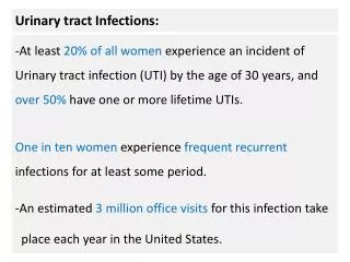
Prevalence of urinary tract infections (UTI) • almost half of all women will have at least one UTI in their lives. • the risk of UTI in women increases after menopause • after a UTI: 20 - 40 % will have a recurrence • recurring infections are usually reinfections. • asymptomatic bacteriuria in women ↑with age & occurs in 2.7% of 15 - 24 year olds 9.3% of over 65 year olds 20 - 50% of over 80 year olds
Prevalence of UTIs 2 • UTI is rare in young and middle-aged men • & often with catheterisation or urological procedures. • bacteriuria in elderly men occurs in • 10% of those living at home, • 20% of those living in nursing homes & • 30% of those who are in hospitals • urinary catheter increases the risk almost ten-fold in hospitalised patients and those in other care homes. • pyelonephritis is common in patients who have been catheterised for over a month.
Gender and sexual contact Infant and children : Male >> female Adolescent-menopause : Female >> male Older age : Female = = male Women: Short ureter Sexual contact : colonization of pathogens in bladder Spermicidal : Change of normal flora Men (<50) : Prostate infection, anatomical defects , Lack of circumcision, homosexuals
Symptomatic Infection Asymptomatic bacteriuria
Urinary Tract Infection- Types urethritis cystitis Lower prostatitis pyelonephritis intrarenal & Upper perinephric abscess
UTI- types by complicating factor • UncomplicatedUTi: infection in a structurally and neurologically normal urinary tract • Complicated UTI: infection in tract with functional or structural abnormalities
Complicated or Un-Complicated UTI • UncomplicatedUTI is the one occurring in a healthy young nonpregnant woman, • & a Complicated UTI is the one occurring in anyone else.
vs Pathophysiology
Host protective factors inUTI • Flushing mechanism of micturition • Acid pH of urine (4.6- 6) anti-bacterial • Acid vaginal pH (3.5-4.5)) suppresses colonization • Urinary Tamm-Horsefall protein (secreted by ascending loop of Henle) & blocks E. coli • Chemotactic factors - interleukin-8
Immune responses in UTI • Submucosal IgA-producing plasma cells in bacterial cystitis • IgM & IgG antibodies produced against bacterial antigens • Protective role of antibodies unclear, may limit damage within the kidney & prevent persistent colonization & thus recurrence of infection
Host Factors Complicating Bacteriuria Residual bladder urine after voiding Turbulent urethral flow(stricture) Foreign bodies Atrophic vaginal mucosa Vesico-ureteral reflux Facilitate bacteriuria Worse prognosis of UTIs Childhood pyelonephritis Diabetic nephropathy Malignant hypertension Chronic pyelonephritis
Bacterial virulence factors in UTI • Escherichia coli strains expressing O-antigens cause high proportion of infections • Capsular antigens of E. coli associated with clinical severity (antiphagocytic) • P-fimbriae enhance attachment of E. coli) to uroepithelial cells • Motile bacteria ascend the ureter against urine flow
Bacterial virulence factors in UTI • Bacterial urease (Proteus) splits urea →NH4 ion alkalinizes urine with loss of acid pH → stone formation → obstruction & survivial of bacteria within stones resisting eradication • Gram-negative endotoxin decreases ureteral peristalsis • Hemolysin damages renal tubular epithelium & promotes invasive infection • Aerobactin of E. coli promote iron accumulation for bacterial replication
Pathogenesis of UTI • Ascending route most common • Colonization of urethra and peri-urethral tissue is the initial event • More in women than men due to short female urethra ,so urethral organisms enter bladder during micturition & in close proximity to perianal areas • Once in the bladder , multiply, then pass up the ureters (esp. if vesico-ureteral reflux) to the renal pelvis & parenchyma • Hospital infection associated with lower urinary tract instrumentation (catheterization, cystoscopy)
Pathogenesis of UTI- blood borne • Hematogenous seeding less frequent than ascending infection • Kidney a common site of abscess formation in Staphylococcus aureusbacteremia, less often in candidemia, rarely with gram-negative bacteremia • Hematogenous seeding of kidney also occurs with Salmonella (typhoid) and Mycobacterium tuberculosis • Source of uropathogens: enteric bacteria
Pathogenesis of UTIs: Risk Factors • ↓resistance of mucous membranes (e.g. in menopause) • sexual intercourse • disturbances in ureteral functioning • in children the re-entering of urine back into the ureters (vesicoureteral reflux), • Uterine prolapse • Diabetes
Pathogenesis of UTIs - 2 Predisposing Conditions- contd • benign prostatic hypertrophy • any illness, eg diabetes, affecting the emptying -Neurogenic Bladder • spinal injury →disturbances in bladder emptying or urinary catheter) • Stones • catheterisation & other urological procedures • Renal Transplantation
Terminology of UTI • Cystitis: localized infection of the bladder • Prostatitis: localized infection of the prostate = lower UTI • Pyelonephritis: infection of the kidney with acute inflammation of the pelvis, medullary and cortical tubules, & intersititum = upper UTI • Urosepsis: bacteremia ( in blood) due to pyelonephritis • Perinephric abscess: associated with obstruction of an infected kidney with abscess formation in the peri-nephric space due to extension of infection across the renal capsule
Urethritis ? • Acute dysuria, frequency • Suspect sexually transmitted pathogens, no hematuria, no suprapubic pain, new sexual partner, cervicitis
Cystitis • Symptoms: frequency, dysuria, urgency, suprapubic pain • Cloudy, malodorous urine (nonspecific) • Leukocyte esterase positive = pyuria • Nitrite positive (but not always) • WBC (2-5 with sx) and bacteria on urine microscopy
Pyelonephritis • Fever, chills • Nausea/Vomiting, diarrhea, tachycardia, • Costo-vertebral angleTenderness or deep abdominal tenderness • Leukocytosis • Urine microscopy: Pyuria + WBC casts, bacteria & hematuria • Possibly signs of Gram negative sepsis
?Pyelonephritis • Complications: sepsis, papillary necrosis, abscess, ureteral obstruction ↓renal function if scarring, in pregnancy – ↑incidence of preterm labor
Diagnosis of UTI • History • Physical exam • Lab parameters: • Urinalysis with microscopy for WBC, bacteria • Urine culture with sensitivities • Diagnose acute uncomplicated cystitis in women based on Hx, PE, and UA alone, no need for culture to treat
Diagnosis • Urinalysis • Leukocyte-esterase positive. = pyuria • Nitrite positive from urease producing bacteria eg proteus (but not always) • Microscopy – WBC ( >5 in pt with symptoms) -- Bacteria
Dipstick Methods pH, Sp.Gravity, protein, glucose, blood, ketone, Leukocyste esterase + Pyuria Nitrite + Gram(-) bacteriuria except pseudomonas
FACTS NITRITE:Screening test for bacteria in urine Gm –ve bact convert urine nitrate to nitrite False –ve: enterococcus, ascorbate, urine <4 hrs LEUCOCYTE – ESTERASE by granulocytes Test threshold is 5 – 15 WBC/HPF False –ve in : glycosuria, high sp. Gravity, cephalexin, tetracyclin, ↑oxalates, vaginal debris
Microscopic Examination Pyuria: WBC > 5 / HPF in spun urine Bacteriuria > 105 cfu/ml
Diagnosis of UTI • Urinalysis : • Leukocyte esterase : On Dipstick exam • Nitrate nitrite : On Dipstick exam • gram negative bacteria except pseudomonas • Pyuria : > 5 WBC /HPF, WBC cast • Bacteriuria : > 105 cfu/ml
Collecting a sample • in adults, mid stream urine (MSU) sample usually reliably represents the urine in the bladder. • samples from urinary bags or bedpans should not be used as they invariably will be contaminated • the most reliable sample is obtained via a suprapubic puncture • urine in bladder >4 hours (any shorter time will increase the risk of false negative findings)
Diagnosis- interpretation • Urine culture • 105 colonies per mL considered standard for diagnosis - but misses up to 50% • Now, 103 to 104 accepted as significant if patient symptomatic • if several bacterial strains are grown on culture; contaminationof the sample is the likely cause • Sensitivities for better tailoring of therapy
Asymptomatic bacteriuria microbiological diagnosis Positive Urine culture repeatedly (105 cfu/ml) in the absence of signs of UTI • pyuria does not affect interpretation • Investige & treat asymptomatic bacteriuria → only in pregnant women Its prevalence varies from 1-5% to 100% in selected population groups. .
Causative agents of UTIs • Escherichia coli • most common • 80% of community - acquired • 50% of hospital-acquired • Others: • enterococci • Staphylococcus saprophyticus and • klebsiella • pseudomonas and proteus are more rare
Localization of UTI No definite standard method Ultrasonography IVP Abdominal CT / MRI Tc-99m DMSA renal scan
A B C D CT images at different levels –> hypodense areas of ?pyelonehritis
T1 MRI- hypointense area T2 MRI- hyperintense area Axial MRI- hyperintense area MRI – Fluid (abscess) in lower pole
Emphysematous Pyelonehritis: (a) CT – air in wall of U.B (b) Plain X ray- mottling in UB (A gas forming infection of renal parenchyma & perinephric tissues )