Download
1 / 1
10 likes | 117 Views
Segmentation of Cerebrovascular Pathologies in Stroke Patients with Spatial and Shape Priors.

E N D
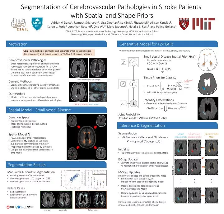
Segmentation of Cerebrovascular Pathologies in Stroke Patients with Spatial and Shape Priors Adrian V. Dalca1, Ramesh Sridharan1, Lisa Cloonan2, Kaitlin M. Fitzpatrick2, Allison Kanakis2, Karen L. Furie3, Jonathan Rosand2, Ona Wu2, Mert Sabuncu4, Natalia S. Rost2, and Polina Golland1 1CSAIL, EECS, Massachusetts Institute of Technology 2Neurology, MGH, Harvard Medical School 3Neurology, RUH, Alpert Medical School. 4Martinos Center, Harvard Medical School Motivation Generative Model for T2-FLAIR Goal: automatically segment and separate small vessel disease (leukoaraiosis) and stroke lesions in T2-FLAIR of stroke patients. We model three tissue classes: small vessel disease, stroke, and healthy • Small Vessel Disease Spatial Prior • Generate parameters • Cerebrovascular Pathologies • Small vessel disease predictor of stroke outcome • Pathologies have similar intensities in T2-FLAIR • Stroke has no consistent shape or location pattern • Clinicians use spatial patterns in small vessel disease to differentiate from stroke lesions • Current Methods • Segment hyperintensities via intensity thresholds • Shape models used for other segmentation tasks • Our Method • Model combines intensity and spatial patterns • Inference to segment and differentiate pathologies • Tissue Priors for Class • Add MRF for spatial contiguity Stroke patients with pathologies small vessel disease stroke healthy • Intensity Observations • Generated independently from Gaussian small vessel disease, stroke lesions Spatial Model - Small Vessel Disease Joint Probability • Common Space • Register training subjects • Maps of small vessel disease overlap(obtained manually) • Spatial Model • PCA on maps of small vessel disease • Components capture co-variation e.g. bilateral periventricular symmetry • Properties match those used by clinicians • Can project estimated small vessel disease onto model Inference & Segmentation • Segmentation • MAP estimate via Variational EM inference • Initialize • Hyperintense voxels: small vessel disease, stroke • E-Step Update • Estimate small vessel disease spatial prior via regularized projection of small vessel disease • M-Step UpdatesSmall vessel disease and stroke probability maps • Estimate for class statistics Include healthy tissue heterogeneity model • Update tissue prior based on previous MAP estimates and • Update posterior using new class statistics, tissue prior, and neighbor agreement • Convergence leads to delineation of small vessel disease and stroke lesions simultaneously Stroke lesion prob. Small vessel disease prob. Spatial model Segmentation Results iterations • Manual vs Automatic segmentation • Good agreement of lesion outlines • Volume agreement (100 subj) • Volume agreement across manual raters • Failure Cases • Bad registration • Large extent of small vessel disease volumes . . . . . . . . . manual automatic failure cases