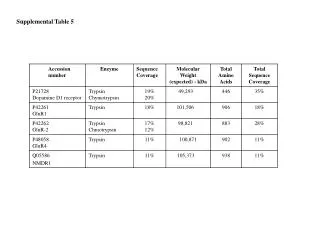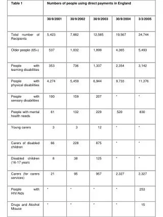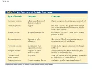
Table 5-1
250 likes | 431 Views
Table 5-1. The enzyme sucrase hydrolyzes its substrate sucrose (a disaccharide). Substrate (sucrose). LE 5-16. Glucose. Enzyme (sucrase). Fructose. a carbon. LE 5-UN78. Carboxyl group. Amino group. LE 5-17a. Alanine (Ala). Valine (Val). Isoleucine (Ile). Glycine (Gly).

Table 5-1
E N D
Presentation Transcript
The enzyme sucrase hydrolyzes its substrate sucrose (a disaccharide) Substrate (sucrose) LE 5-16 Glucose Enzyme (sucrase) Fructose
a carbon LE 5-UN78 Carboxyl group Amino group
LE 5-17a Alanine (Ala) Valine (Val) Isoleucine (Ile) Glycine (Gly) Leucine (Leu) Nonpolar Methionine (Met) Phenylalanine (Phe) Proline (Pro) Tryptophan (Trp)
LE 5-17b Polar Tyrosine (Tyr) Serine (Ser) Asparagine (Asn) Threonine (Thr) Cysteine (Cys) Glutamine (Gln)
LE 5-17c Acidic Basic Electrically charged Aspartic acid (Asp) Lysine (Lys) Arginine (Arg) Glutamic acid (Glu) Histidine (His)
Peptide bond LE 5-18 Side chains Peptide bond Backbone Carboxyl end (C-terminus) Amino acid (N-terminus)
LE 5-20 Hierarchical organization of protein structures b pleated sheet +H3N Amino end Amino acid subunits helix Primary Secondary Tertiary Quaternary
Primary Structure Fig. 5-21a 1 5 +H3N Amino end 10 Amino acid subunits 15 20 25
Secondary Structure Fig. 5-21c pleated sheet Examples of amino acid subunits helix
Tertiary Structure Hydrophobic interactions and van der Waals interactions Polypeptide backbone Hydrogen bond Disulfide bridge Ionic bond LE 5-21d
Hydrophobic interactions and van der Waals interactions LE 5-21f Polypeptide backbone Hydrogen bond Disulfide bridge Ionic bond
Fig. 5-21e Tertiary Structure Quaternary Structure
Polypeptide chain LE 5-21g b Chains Iron Heme a Chains Hemoglobin Protein complex Collagen
LE 5-19a Groove A ribbon model
LE 5-19b Groove A space-filling model
Biology and art… http://www.julianvossandreae.com/
Denaturation LE 5-23 Normal protein Denatured protein Renaturation
LE 5-24a X-ray diffraction pattern Photographic film Diffracted X-rays X-ray source X-ray beam Crystal
Nucleic acid Protein LE 5-24b X-ray diffraction pattern 3D computer model
5¢ end LE 5-26a Nucleoside Nitrogenous base Phosphate group Pentose sugar Nucleotide 3¢ end Polynucleotide, or nucleic acid
Nitrogenous bases Pyrimidines LE 5-26b Uracil (in RNA) U Cytosine C Thymine (in DNA) T Purines Guanine G Adenine A Pentose sugars Deoxyribose (in DNA) Ribose (in RNA) Nucleoside components
5¢ end 3¢ end Sugar-phosphate backbone LE 5-27 Base pair (joined by hydrogen bonding) Old strands Nucleotide about to be added to a new strand 5¢ end New strands 5¢ end 3¢ end 5¢ end 3¢ end
DNA LE 5-25 Synthesis of mRNA in the nucleus mRNA NUCLEUS CYTOPLASM mRNA Movement of mRNA into cytoplasm via nuclear pore Ribosome Synthesis of protein Amino acids Polypeptide