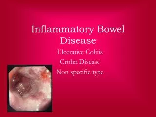
Bowel Disease
1.25k likes | 1.45k Views
Mark Bromley Emergency Medicine PGY-3. Bowel Disease. large and Small. Overview. Cases Approach – Work-up Appendicitis Dealing with surgeons Mesenteric Ischemia ABD films SBO. Case. 21 ♂ with ABD pain onset ~ 24h Pain Peri -umbilical Escalating to 8/10 Fevers/Chills

Bowel Disease
E N D
Presentation Transcript
Mark Bromley Emergency Medicine PGY-3 Bowel Disease large and Small
Overview • Cases • Approach – Work-up • Appendicitis • Dealing with surgeons • Mesenteric Ischemia • ABD films • SBO
Case 21 ♂ with ABD pain onset ~ 24h • Pain • Peri-umbilical • Escalating to 8/10 • Fevers/Chills • Emesis x 3 this AM OE: • 38.1oC 16 85 122/81 • ABD: • Diffuse peri-umbilical • No Rebound/Guard
Case PMHx: Well PSHx: None Meds: Nil Allergies: NKDA
Approach • Anatomic • Systems
Work-up • Bloodwork • CBC, lytes, Cr • INR, PTT • ALT,ALP,Bili, GGT • Lipase • Blood cultures • ♀ BHCG • ESR/CRP • Urine R&M • Imaging • Plain films - 3 views • CT ABD/Pelvis • US
Case • Order your work-up • Morphine 2.5-5mg IV for pain • Return in 1-2h • Comfortable • Pain – now in RLQ • Tender at McBurneys
Appendicitis - Classic • Pain • Vague peri-umbilical pain that localizes to the RLQ (McBurney’s) • …↑ over 12 to 24h period • Pain lasting more than 36h is rare – or perfed • Febrile • Anorexic • Elevated WBC • Rosvings, Psoas, Obturator
Signs • Psoas sign • With pt supine, flex hip against resistance by pushing down against knee -- pain = +ve • Obturator sign • Passively flex hip & knee and internally rotate leg at the hip -- pain = +ve • Rosvings sign • press down in LLQ then release suddenly - pain = + ve
Differentiate into 3 groups • High suspicion for appendicitis – need for immediate surgery • i.e. classic presentation • Intermediate suspicion for appendicitis – no clear-cut need to go to OR yet • Atypical presentation • Low suspicion for appendicitis
Appendicitis • Expedient diagnosis • Non-ruptured - - - - - - - - - - - Mortality 0.6% • Ruptured - - - - - - - - - - - - - - Mortality 5% …the blood was clotted …nurses are sure the lab dropped it • Surgeon wants a WBC before seeing
Appendicitis – Role of WBC • Methods: • prospective consecutive case series • All patients presenting to the ED in whom the diagnosis of appendicitis was the attending physician’s primary consideration • Patient temperature as taken in the ED, initial total WBC count, and discharge diagnosis. • Results: • N=293 • wbc > 10 (+LR) 1.59 (-LR) 0.46 • wbc > 12 (+LR) 2.70 • Fever > 37.2oC (+LR) 1.30 (-LR) 0.82
Liklihood Ratio • likelihood ratio, is the ratio of the maximum probability of a result under two different hypotheses Probability of ↑WBC with Appy ---------------------------------------- = LR Probability of ↑WBC w/o Appy
Probability of ↑WBC with Appy ____________________ Probability of ↑WBC w/o Appy
Design: • Random assignment of vignettes with different presentation formats of diagnostic test accuracy. • Setting: • Auditorium at a continuing medical education conference. • Participants: 183 physicians. • Intervention: • After estimating probabilities of 6 common illnesses described in patient vignettes, physicians • Results: • post-test probability estimates deviated to a small and similar extent from Bayes-based • estimates in the groups informed by sensitivity and specificity or likelihood ratios. • An inexact numerical graphic led physicians to come closer to Bayes-based estimates in the PE and chronic obstructive pulmonary COPD vignettes • some physicians estimated lower illness probabilities after a positive test result if it was accompanied by a low test accuracy value.
Odds and Probability …only works with odds I’ll give you twenty to one odds 20:1 probability = 20/total (21) = 95% chance Forty to sixty odds = 40:60 = 40/60 = 0.66 probability = 40/total (100) = 40% chance
Likelihood Ratio • % chance this guy has an appy = 0.4 (40%) • Convert that to odds (pretest) • 0.4/0.6 = 4/6 = 2/3 • (2/3) x 3 = 6/3 = 2 • Convert back to probability (posttest) • 2/3 = 0.67 (67%)
Likelihood Ratio • As a rule • (+) LR > 10 • (-) LR <0.1 …useful
Case • Resident comes down and sees the patient …hmm, didn’t do a rectal? Wow. hmmm….
Appendicitis - Rectal • Why do we do a rectal exam? • Should we do a rectal exam? • Looking for other diagnoses • PR bleeding • Peri-anal disease • Mass in the vault • Does everyone need a rectal?
Patients and Methods: • 100 consecutive adults admitted to the emergency surgical unit with acute abdominal pain • Following DRE, patients completed an anonymous questionnaire • The house officer conducted the rectal examination at admission and also completed an evaluation sheet • Results: • A working diagnosis of acute appendicitis in 38 patients and gastroduodenal, pancreatobiliary pathology in 24 patients was made • DRE did not alter clinical diagnosis or initial management in any patients • Routine DRE did not detect any unrelated pathology • 93 wanted to know why rectal examination was required • 78 patients rated the DRE as uncomfortable • 43 were willing for DRE as a routine • 54 patients preferred to have the DRE at the time of other bowel tests rather than at emergency admission
Patients • 1204 consecutive patients admitted to hospital with RLQ pain • 1028 had a rectal examination on admission • Main outcome measures - Odds ratio for each symptom and sign related to final diagnosis • Results of multiple logistic regression analysis for acute appendicitis • Results • Right sided rectal tenderness (odds ratio 1.34, p<005) • RLQ tenderness (odds ratio 5.09) • Rebound tenderness (3.34) • Guarding (3.07) • Muscular rigidity in the abdomen (5.03) • In the logistic regression analysis of patients with acute appendicitis, when allowance was made for the presence or absence of rebound tenderness, rectal tenderness on the right lost its significance • Six patients had masses palpable rectally, of which three were palpable on abdominal examination; the other three patients had acute appendicitis. • No other unexpected diagnoses were established, and no useful additional • Conclusion • If patients presenting with pain in the RLQ of the abdomen are tested for rebound tenderness then rectal examination does not give any further diagnostic information
Case The resident agrees – this sure looks like appendicitis. But the boss would like some imaging. …thoughts?
Ultrasound (Graded Compression) • Test Characteristics • Sensitivity 75-90%, Specificity 86-100% • Pros • No radiation, safe in kids, pregnant pts • Can identify alternate Dx esp. in female pts • Cons • Difficult for us to get • Operator-dependant • Limited in obese pts or with ↑ bowel gas • Identifies alternate Dx less often than CT • Painful
CT scan • Test characteristics • Sensitivity 90-100%, specificity 91-99% • Pros • Identifies alternate Dx more often than U/S • Fast & accessible in our practice setting • Cons • Radiation dose (~100 CXR’s) • Delay time to surgery • Multiple techniques in literature: controversial as to which is best but all ~90-100% sensitive • Less accurate in pts w/ little intraabdominal fat
CT vs U/S • 2 prospective RCT’s of U/S vs CT • CT more sensitive & specific than U/S • 94-97% sensitive vs. 76 – 100% for U/S • 100% specificity vs. 76-90% for U/S • More alternate Dx identified by CT Horton et al. Am J Surg 2000; 179: 379-81 Walker et al. Am J Surg 2000; 180: 450-55
CT vs U/S Methods: • 120 consecutive pts 8-81 yo w/ ?appy who were too well to go to OR but too ill to simply D/C • Did focused CT w/ rectal contrast & U/S w/in 1 hr • Gold standard - pathology or clinical f/u x 6m Results: • CT: 95% sensitive, 89% specific • U/S: 87% sensitive, 74% specific • CT identified 14 alternate Dx vs. 9 for U/S • U/S missed 2/3 of pts w/ perforation *Pickuth et al. Suspected acute appendicitis: Is ultrasonography or computed tomography the preferred imaging technique? Eur J Surg. 2000; 166: 315-19
Does imaging change mgmt? • 2 studies of CT in pts w/ ? appendicitis comparing Tx plan before & after access to results of scans Results: • CT changed disposition in 27 – 59% of pts • Prevented d/c of ~3% pts w/ appendicitis • Prevented negative laparotomy in 3-13% • Alternate Dx in 11-20% • …yes • Frank et al. Unenhanced helical CT scanning of the abdomen and pelvis changes disposition of patients presenting to the emergency department with possible acute appendicitis. J Emerg Med 2002; 23: 1-7 • Rao et al. Effect of computed tomography of the appendix on treatment of patients and use of hospital resources. N Eng J Med. 1998; 338: 141-6
Bottom line • Group 1 • Appendectomy regardless of imaging result • Group 2 • Image • Group 3 • Clearly instructed when to return for re-evaluation
Appendicitis - Mgmt • Hydration • Antibiotics • Ancef/Flagyl (surgical wound) • Fluroquinalone/Flagyl (gram(-) rods / anaerobes) • Surgery
Case • 65 ♂ with ABD pain • Diffuse ABD pain 8/10 • Rapid onset • Opiod resistant • N/V/D • Watery stools x 3 • OE: • 104 20 145/67 37.2 • ABD: Diffuse tenderness - no rebound/guard • Rectal: Normal (-) FOB • PMHx: • HTN/DMII/smoke/AFIB/MI x3
Approach • Differential
Ischemic bowel - etiology • Embolic • LA • LV • Cardiac Valves • SMA is most susceptible to embolism • Multiple emboli • Concomitant vasoconstriction occurs
Ischemic bowel - etiology Thrombotic • Arterial • Acute event • Chronic intestinal ischemia from progressive atherosclerosis • Involves multiple vessels • Venous • venous thrombosis →mesenteric venous flow → bowel wall edema, fluid efflux into lumen • ↓BP • ↑ blood viscosity • Risk Factors • Hypercoagulable states • Portal hypertension • Abdominal infections • Blunt abdominal trauma • Pancreatitis • Splenectomy • Malignancy in the portal region
Ischemic bowel - etiology • Non-occlusive etiology • systemic illness → systemic shock → ↓CO • cocaine → vasospasm • Venous thrombosis → ↓ venous return → interstitial swelling of bowel wall → ↓ arterial flow
Mesenteric Ischemia – clinical • ABD pain • rapid onset • severe • out of proportion to exam • N/V/D • forceful bowel evacuation • Risk factors • AFIB • CHF • peripheral vascular disease • hypercoagulability
Ischemic bowel - diagnostics • Labs (non-specific) • Lactate • WBC • INR/PTT • Imaging • Plain films (nonspecific late findings – not useful) • Thumbprinting • Pneumatosisintestinalis • Portal venous gas • CT • Angiography
CT scan • sensitivity 64-100% • specificity 89-94% • Evidence of ischemia in bowel wall & mesentery • Evidence of clot in SMA • First investigation done routinely here • If suspect mesenteric ischemia let radiology know • Good but not good enough • If CT is negative & high pre-test probability you need an angiogram
Ultrasound • Doppler can determine major obstruction to flow in venous & arterial systems • Dilated, tubular vessels with echogenic material (clot) • Abnormal flow • Limitations • Studied primarily in venous thrombosis & chronic mesenteric ischemia • Unsure how it performs for acute mesenteric ischemia • Only good for more proximal blockages • Has limitations inherent to all U/S exams
Angiography • Gold standard (~90% sens) • Diagnostic and therapeutic • Infuse vasodilators into SMA (papaverine) • Angioplasty • Drawbacks • Time-consuming • Risks of contrast & invasive procedure • Expensive
Angiography: Early vs Late • Angiography → early in pts w/o peritonitis & ↑ suspicion • Can buy time (papaverine) • Can aid in surgical decision making • Surgical: embolectomy, thrombectomy, endarterectomy, bypass graft • Non-surgical: angioplasty • Early (before peritonitis) angio & intervention • ↓ mortality 70-90% → 10% • Down side: • ↑ negative angios • Associated risks & costs
Angiography: When to say no • Contraindications: • Unstable hypotensive pts on vasopressors • Difficult to differentiate b/w occlusive & non-occlusive etiologies • Can’t infuse vasodilators • Pts w/ peritonitis • Delays surgery
Case • 32 ♀ with nausea and vomiting • Abdominal pain • periumbilical and crampy • paroxysms of pain q 4-5 min • abdominal distension • Vomiting q 30 min • BM none x 48h • PMHx • Crohns – dx in 1997 - resection 2002, 2007
Crohn’s Extra-intestinal manifestations • Skin manifestations • erythemanodosum, • pyodermagangrenosum • Peripheral arthritis (asymmetric involvement of larger joints) • Ankylosingspondylitis and sacroiliitis • Aphthous ulcers • Ocular manifestations (eg, episcleritis, recurrent iritis, uveitis) • Amyloidosis and thromboembolic manifestations • Liver • elevation of enzyme levels • Cholangitis • Autoimmune chronic active hepatitis, and cirrhosis
Comparisons of Crohn's and UC Crohn'sUlcerative colitis Terminal ileum involved Commonly Seldom Colon involvement sually Always Rectum involvement Seldom Usually Peri-anal disease Common Seldom Bile duct involvement No ↑ in PSC Higher rate Distribution of Disease Patchy (Skip lesions) Continuous Endoscopy Deep geographic ulcers Continuous ulcer Depth of inflammation May be transmural Shallow, mucosal Fistulae Common Seldom Stenosis Common Seldom Surgical cure Often Cured by colectomy Smoking risk for smokers ↓risk for smokers