Download
1 / 33
330 likes | 619 Views
Evolution of Parathyroid Surgery Using Sestamibi Imaging Guidance. David R. Byrd, MD Department of Surgery University of Washington. Disorders of Parathyroid Glands.

E N D
Evolution of Parathyroid Surgery Using Sestamibi Imaging Guidance David R. Byrd, MD Department of Surgery University of Washington
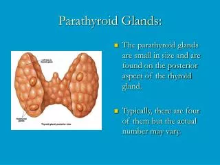
Disorders of Parathyroid Glands • hypoparathyroidism -rare. Almost always caused by excessive surgical removal of parathyroid tissue (iatrogenic) during thyroid or parathyroid surgery • hyperparathyroidism (HPT): • primary - hi Ca++, hi PTH - usually due to single adenoma (90%), cured by removal of adenoma • secondary - lo Ca++, hi PTH, seen in chronic renal failure - not a surgical problem • tertiary - hi Ca++, hi PTH, seen after renal transplant - hyperplasia of all 4 glands
Traditional Surgery for Hyperparathyroidism • primary HPT - 4 gland exploration, remove adenoma, biopsy 3+ normal glands • tertiary HPT (after renal transplantation) - 3 1/2 gland removal +/- forearm autotransplant
Complications of Parathyroid Surgery • persistent HPT - 1-20% (experience dependent) • temporary or permanent hypocalcemia - 1-20% • nerve injury - recurrent or superior laryngeal -1-10% • bleeding - <5%
Unilateral Exploration for Primary HPT • if: one abnormal, hypercellular gland and one normal gland found on one side, no contralateral exploration • occasional use of preop thallium-technetium scan • results of 5 studies - cure 93-100%
Indications for Operation in Asymptomatic Patient w/ Primary HPT - NIH Consensus(1990) • markedly elevated serum Ca++ • episode of life-threatening hyperCa++ • reduced creatinine clearance • renal stones • markedly elevated 24 hr urinary Ca++ • substantially reduced bone mass (by DEXA scan) • age <50 (relative indication for surgery)
Parathyroid Imaging • Tc-99m sestamibi scan (Cardiolyte) • ultrasound • initially thought useful only in persistent or recurrent disease • thallium-technetium subtraction scan - now rarely used
Tc-99m Sestamibi Scan • taken up by actively metabolizing tissues - salivary glands, thyroid, parathyroid glands • over time, blood flow causes washout from thyroid and normal parathyroid glands • delayed images show a discrete “hot spot” in 75-80% patients with primary HPT • can be used to direct minimally invasive surgical approaches
Parathyroid Imaging -Tc-99m Sestamibi 45 min Anterior 45 min LAO submandibular gland thyroid lobe adenoma Delayed views 2 HR 2 HR
Right inferior pole parathyroid adenoma 15 min Ant 1 hr Ant 1 hr RAO adenoma
Right superior parathyroid adenoma 15 min Ant 1 hr Ant adenoma
Advances Enabling Localized Exploration • Tc-99m sestamibi radioguided exploration • rapid IOPTH assay - 1/2 life = 3-5 minutes
Rapid IOPTH Assay • exploits short half life (3-5 minutes) of PTH • serum baseline level #1 prior to exploration • level #2 after exploration but before removal adenoma • levels 5 & 10 minutes after adenoma removal • 5 minute level > 50% second baseline level = high prediction of success -Irvin G, et al, 1993
Studies of IOPTH Measurement in HPT solitary/ Uni/bilat. Cure rate # pts MGD exploration ( %) Nussbaum 1988 12 12/0 8/4 100 Chapuis 1996 173 -- 160/13 94 Irvin 1993 61 -- -- 90 Sofferman 1998 40 31/9 -- 100 Carty 1997 67 58/9 42/25 99 Irvin 1994 18 18/0 -- 89 Starr 2001 50 38/12 0/50 92
Minimally Invasive Radioguided Parathyroidectomy (MIRP) • only in patients who localize by pre-op sestamibi scan (75% with primary HPT) • sestamibi scan performed 2-3 hours before exploration - timing crucial • gamma probe used to find the “hottest” spot • ex vivo adenoma counts >20% background • no further dissection and no frozen section • if no adenoma found, 4 gland exploration -Norman J, et al, 1997
MIRP - results • 2 cm incision • local w/ sedation, out-patient procedure • 100% cure rate • no complications • mean operating time = 25 minutes • re-operative cure rate = 100% -Norman J, 1997
Studies of MIRP in HPT solitary/ Uni/bilat. Cure rate # pts MGD exploration ( %) Martinez 3 2/1 -- -- Gallowitsch 12 -- -- -- Bonjer 62 49/10 -- 95 Norman 15 15/0 14/1 -- Norman 24 21/0 21/1 -- Flynn 39 32/6 30/9 100
Evolution of Surgery for Primary HPT • Preoperative sestamibi in all patients with primary HPT: • help decision whether to operate in selected patients • localize adenoma to plan localized exploration • Minimally invasive parathyroidectomy (MIP): • 2-4 cm incision • often w/ local + sedation • out-patient procedure • +/- IOPTH testing - biochemical confirmation • Endoscopic removal of parathyroid gland(s)
Right inferior parathyroid adenoma - 54F 15 min Ant 1 hr Ant 1 hr RAO adenoma
IOPTH Testing and Results MIP findings - 500mg L inferior pole adenoma Baseline #1 214 Baseline #2 157 5 minute post 32 10 minutes post 20 F/U levels 3 mos: Ca++ = 9.5, PTH = 55 (both normal)
Case # 3 50M, asymptomatic: - serum Ca++ = 13.4 - preop iPTH = 750 - concern for carcinoma
Tc-99m sestamibi positive for intense uptake LIP Delay Ant Immed Ant
IOPTH Testing and Results Case #3: 50M, 4.2 LIP gm adenoma Baseline #1 1259 Baseline #2 764 5 minute post 129 10 minutes post 93 Early F/U: Ca++ =8.8, PTH = 138 (low calcium, sl. elevated PTH)
Operation for Tertiary HPT • standard operation remains 3 1/2 gland removal or total parathyroidectomy w/ auto transplant dorsal forearm • Imaging not standard at present • selected patients may benefit from Tc-99m sestamibi preop scan • role of IOPTH testing evolving