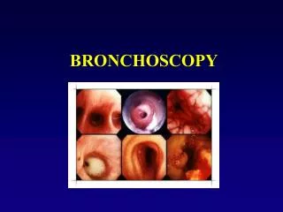

Indication for diagnostic bronchoscopy
E N D
Presentation Transcript
1. 1
2. 2
3. 3 GUIDELINES ON DIAGNOSTIC FLEXIBLE BRONCHOSCOPY
4. 4 THE AREAS COVERED These guidelines have been developed by the British Thoracic Society (BTS) and published in thorax 2001,56;(suppl.1)
The areas covered by these guidelines are as follows:
Complications, Contraindications and Precautions;
Sedation and Anaesthesia/analgesia;
Cleaning and Disinfection Including Glutaraldehyde Usage;
Staff Safety;
Bronchoscopy in the Intensive Care Unit;
Data Collection and Staff Training;
Patient Satisfaction.
5. 5 PATIENT SAFETY
6. 6 BEFORE BRONCHOSCOPY(1) Patients with suspected chronic obstructive pulmonary disease (COPD) should have spirometric parameters checked before bronchoscopy and, if the COPD is found to be severe (FEV1 <40% predicted and/or SaO2 <93%), should also have arterial blood gas tensions measured.
Oxygen supplementation and/or intravenous sedation may lead to an increase in the arterial CO2 level and hence sedation should be avoided where the pre-bronchoscopy arterial CO2 is raised and oxygen supplementation given only with extreme caution.
Prophylactic antibiotics should be given before bronchoscopy to patients who are asplenic, have a heart valve prosthesis, or a previous history of endocarditis.
Bronchoscopy should be avoided if possible within 6 weeks of a myocardial infarction.
7. 7 BEFORE BRONCHOSCOPY(2) Verbal and written patient information improves tolerance of the procedure by the patient and should be provided.
Asthmatic subjects should be premedicated with a bronchodilator before bronchoscopy.
Routine preoperative checks of the platelet count and/or prothrombin time are only required in those patients with known risk factors having routine bronchoscopy without transbronchial biopsy.
If it is anticipated that biopsy specimens may be required at bronchoscopy, oral anticoagulants should be stopped at least 3 days before bronchoscopy or they should be reversed with low dose vitamin K.
On the rare occasions when it is necessary to continue with anticoagulants, the international normalised ratio (INR) should be reduced to <2.5 and heparin should be started
8. 8 BEFORE BRONCHOSCOPY(3) Platelet count, prothrombin time, and partial thromboplastin time should be checked before performing transbronchial biopsies.
It is suffcient for patients to have no food by mouth for 4 hours and to allow clear fluids by mouth up to 2 hours before bronchoscopy.
Intravenous access should be established in all patients before bronchoscopy is commenced (and before sedation, if given) and left in place until the end of the postoperative recovery period.
Sedation should be offered to patients where there is no contraindication.
Atropine is not required routinely before bronchoscopy
9. 9 DURING BRONCHOSCOPY (1) Patients should be monitored by oximetry.
Oxygen supplementation should be used to achieve an oxygen saturation of at least 90% to reduce the risk of significant arrhythmias during the procedure and also in the postoperative recovery period.
The total dose of lignocaine (lidocaine) should be limited to 8.2 mg/kg in adults (approximately 29 ml of a 2% solution for a 70 kg patient) with extra care in the elderly or those with liver or cardiac impairment.
Lignocaine gel (2%) is preferred to lignocaine spray for nasal anaesthesia.
The minimum amount of lignocaine necessary should be used when instilled through the bronchoscope.
10. 10 DURING BRONCHOSCOPY (2) Sedatives should be used in incremental doses to achieve adequate sedation and amnesia.
Fluoroscopic screening is not required routinely during transbronchial biopsy in patients with diffuse lung diseases, but should be considered in those with localised lung lesions.
At least two endoscopy assistants should be available at bronchoscopy, and at least one of these should be a qualified nurse.
Routine ECG monitoring during bronchoscopy is not required but should be considered in those patients with a history of severe cardiac disease and those who have hypoxia despite oxygen supplementation.
Resuscitation equipment should be readily available.
11. 11 AFTER BRONCHOSCOPY Postoperative oxygen supplementation may be required in some patients, particularly those with impaired lung function and those who have been sedated.
A chest radiograph should be carried out at least 1 hour after transbronchial biopsy to exclude a pneumothorax
Patients who have had transbronchial biopsies should be given verbal and written advice about the possibility of developing a pneumothorax after leaving hospital.
Patients who have been sedated should be advised verbally and in writing not to drive, sign legally binding documents, or operate machinery for 24 hours after the procedure.
It is preferable that day case patients who have been sedated should be accompanied home and that higher risk patients such as the elderly or those from whom transbronchial biopsy specimens have been taken should have someone to stay with them at home overnight.
12. 12 BRONCHOSCOPE CLEANING AND DISINFECTION (1) Compatibility of decontamination methods should be checked with the manufacturers of bronchoscopic instruments and accessories.
Decontamination and disinfection should be carried out at the beginning and end of a list and between patients.
Cleaning and disinfection of bronchoscopes should be undertaken by trained staff in a dedicated room.
Thorough cleaning with detergent remains the most important initial stage of the process.
When 2% glutaraldehyde is used for manual and automated disinfection, immersion for 20 minutes is recommended for bronchoscopes at the beginning and end of a session and between patients.
13. 13 BRONCHOSCOPE CLEANING AND DISINFECTION (2) Longer immersion times of 60 minutes are recommended for known or suspected atypical mycobacterial infections and patients known to be HIV positive.
Patients with suspected tuberculosis should undergo bronchoscopy at the end of the list.
Glutaraldehyde, although widely used for endoscopes, is only slowly effective against mycobacteria. Peracetic acid, chlorine dioxide, and superoxidised water are more rapidly effective (within 5 minutes or less) but are more damaging to instruments and processing equipment, are less stable, and are more expensive. They may, however, be less irritant than glutaraldehyde.
14. 14 STAFF SAFETY
15. 15 STAFF SAFETY(1) All staff should be vaccinated against hepatitis B and tuberculosis, and immunity and tuberculin status should be checked as appropriate.
During bronchoscopy staff should wear protective clothing (gowns or plastic aprons, masks/visors, and gloves).
+ High grade particulate masks should be worn when patients known to have multidrug resistant tuberculosis or those at high risk undergo bronchoscopy and the procedure should be carried out in a negative pressure facility.
Non-powdered latex or non-latex gloves should be worn instead of powdered latex gloves to minimize the risks of latex sensitization to staff and patients
Injection needles should not be re-sheathed, and spiked biopsy forceps require careful cleaning.
16. 16 STAFF SAFETY(2) Bronchoscopes should be disinfected ideally in a dedicated room using well ventilated automated systems, preferably inside a fume cabinet, to prevent unnecessary exposure to disinfectants.
During cleaning and disinfection staff need to wear protective clothing (nitrile gloves and plastic aprons with eye and respiratory protection, where appropriate) to protect them from splashes, aerosols, and vapour.
The use of disposable accessories, especially injection needles, may reduce the risk of infection which may occur during the cleaning of equipment.
Wherever possible, autoclavable or disposable accessories should be used to prevent unnecessary exposure to disinfectants.
Bronchoscopy staff need to be trained in patient care, infection control, and instrument decontamination including the safe use of aldehydes and the potential health risks.
17. 17 STANDARDS AND PERFORMANCE OF DIAGNOSTIC TECHNIQUES At least five bronchial biopsy specimens should be taken in cases of suspected bronchial malignancy.
Biopsies, brushings and washings should all be obtained in cases of suspected endobronchial malignancy.
A minimum diagnostic level of at least 80% should be obtained from a combination of biopsies, brushings, and washings in cases of endoscopically visible malignancy.
When taking transbronchial lung biopsy specimens in patients with diffuse lung disease, an attempt should be made to obtain 4�6 samples from one lung.
18. 18 PATIENT CARE Verbal and written patient information improves tolerance of the procedure and should be provided.
It is suffcient for patients to have no food by mouth for 4 hours and to allow clear fluids by mouth up to 2 hours before bronchoscopy.
Patients who have been sedated should be advised not to drive, sign legally binding documents, or operate machinery for 24 hours after the procedure
19. 19 SEDATION DURING FLEXIBLE BRONCHOSCOPY(1) Are sedatives necessary?
The purpose of sedation is to improve patient comfort for what can be an unpleasant procedure.
Sedation may also make the procedure easier for the bronchoscopist to perform and the patient more willing to accept a repeat procedure (if necessary).
Sedation should benefit the patient who is particularly anxious
sedation should be avoided or used with extreme caution in patients such as those with severe COPD who have an increased risk of responding adversely
20. 20 SEDATION DURING FLEXIBLE BRONCHOSCOPY(2) How sedation is given?
most sedation regimens are based upon a single dose or incremental
doses of an intravenous sedative agent administered at the time of bronchoscopy.
MIDAZOLAM (Dormicum)
Midazolam is a water soluble benzodiazepine with an elimination half life of about 2 hours and is generally preferred to diazepam.
Its onset is rapid and duration of action brief in healthy individuals.
A better approach of giving is incremental dosing which achieves improved tolerance of bronchoscopy,
21. 21 SEDATION DURING FLEXIBLE BRONCHOSCOPY(3) COMBINATIONS WITH NARCOTIC DRUGS
A combination of a benzodiazepine and narcotic has been widely used.
Unfortunately, such a combination may be associated with more arterial desaturation and CO2 retention than when using midazolam alone.
22. 22 FLEXIBLE BRONCHOSCOPY IN THE INTENSIVE CARE UNIT (ICU)
23. 23 FLEXIBLE BRONCHOSCOPY IN (ICU) The internal diameter of the endotracheal tube, through which the bronchoscope is inserted, must be taken into consideration before bronchoscopy.
Intensive care units should have the facility to perform urgent and timely flexible bronchoscopy for a range of therapeutic and diagnostic indications.
Patients in ICU should be considered at high risk from complications when undergoing fibreoptic bronchoscopy.
Continuous multi-modal physiological monitoring must be continued during and after fibreoptic bronchoscopy.
Care must be exercised to ensure adequate ventilation and oxygenation is maintained during fibreoptic bronchoscopy via an endotracheal tube.
More profound levels of sedation/anaesthesia can be achieved in ventilated patients provided the clinician performing the procedure is acquainted with the use of sedative/anaesthetic agents.
24. 24 ENDOTRACHEAL TUBE SIZE The internal diameter of the tracheal tube relative to the external diameter of the bronchoscope is an important consideration.
Bronchoscopes in the non-intubated patient occupy only 10�15% of the cross sectional area of the trachea.
In contrast, a 5.7 mm bronchoscope. occupies 40% of a 9 mm endotracheal tube
and 66% of a 7 mm tracheal tube.
Failure to recognise this may lead to inadequate ventilation of the patient and impaction of or damage to the bronchoscope.
Tracheostomy tubes are also prone to damage the bronchoscope, particularly during withdrawal when the rigid edge of the end of the tracheostomy tube can abrade the covering of the bronchoscope.
Lubrication is essential to facilitate passage of the bronchoscope.
25. 25 VENTILATOR SETTINGS Pre-oxygenation should be achieved by increasing the inspired oxygen concentration to 100%. 100% oxygen should be given during bronchoscopy and in the immediate recovery period.
The ventilator should be adjusted to a mandatory setting. Triggered modes such as pressure support or assist control will not reliably maintain ventilation during fibreoptic bronchoscopy.
A special swivel connector (Portex, Hythe, UK) with a perforated diaphragm, through which the bronchoscope can be inserted and allows continued ventilation.
26. 26 TRAINING(1) Flexible bronchoscopy is a complex and potentially hazardous procedure requiring trained personnel (medical, nursing, and paramedical) to minimise the risk to both patient and staff.�
The optimal number of procedures which should be undertaken under direct supervision (trainer in bronchoscopy unit) and indirect supervision (trainer able to assist if called) before undertaking bronchoscopy alone will vary, depending on the competency of the trainee and the complexity of the procedure being undertaken.
27. 27 TRAINING(2) It would seem reasonable to undertake a minimum of 50 procedures under direct supervision and a further 50 under indirect supervision, although the trainer or other competent bronchoscopist should be available to give advice if needed for any trainee bronchoscopist