Download
1 / 40
400 likes | 416 Views
This chapter explores the study of bacteria, starting with Robert Hooke and Anton van Leeuwenhoek. It delves into the fundamentals of light microscopy, including magnification and resolution, and introduces different types of microscopy techniques.

E N D
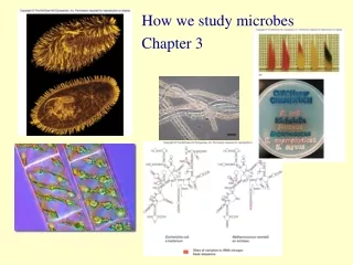
How we study microbes Chapter 3
Bacteria were the first microorganisms to be studied Small in size 1000 X magnification Most about 200 nm to 1 mm • Robert Hooke • Used the term “cell” • 1665 • Anton van Leeuwenhoek • First to visualize bacteria & protozoa • 1678 • Beginning of the end of “spontaneous generation”
Fundamentals of Light Microscopy • Ultimate goal is to obtain information • Two factors affect amount of attainable information • Magnification • Resolution
Magnification - How Does a Lens Magnify? Air and glass have different refractive indices, so light refracts (bends) as it passes through a lens. Image is magnified as once parallel rays now diverge. Virtual Image Reaches the eye E E
Pathway of Light in Compound Microscope Virtual Image formed by ocular lens Mistake- Blue and red should be reversed here (page 75) Magnified primary image This lens not for magnification- concentrates light
Magnification Defined as amount of enlargement of specimen Picture of Anton van Leeuwenhoek 1X 5X 20X 100X • Magnification can be infinite • At some point, no more information can be gathered • Have reached the limiting factor of microscopy - RESOLUTION
Resolution • Minimum distance at which two objects can be • distinguished (the smaller the number, the better the resolution) • Several factors affect resolution • Wavelength (l) of light • Refractive index (h) of medium between lens and • specimen • Distance between lens and specimen • Contrast
l = wavelength of light h = refractive index q = angle between most divergent light ray gathered by lens and the center of the lens l 2hsinq Resolution = Numerical Aperture Mathematical constant that describes the efficiency of a lens 0.1 in a lowest powered lens (4 X) 1.25 in the highest power lens (100 X) Higher numerical aperture =Better Resolution
l 2 N.A. Resolution = l of Light Used l = wavelength of light 500 nm ---------- = 200 nm 2 x 1.25 • Different colors of light have different l • The shorter the wavelength, the higher the resolution- • 400-500 nm is excellent • Thus many light microscopes use filters to select color of light • blue-green l
Refractive Index of Medium Between Specimen and Lens Light path without oil Light path with oil l 2hsinq Resolution= h = refractive index • Refractive index - ability to bend light • Glass from the slide bends light more than air • Light is bent away from lens as it passes through specimen • Immersion oil has similar refractive index as glass • More light is gathered by the lens, more information
Oil Immersion Lens • = 1 for air • = 1.5 for glass • = 1.52 immersion oil
The Distance Between the Lens and the Specimen • The further the lens from the • sample, the more light is lost • The closer the lens, more light • gathered • More light means more • information and higher resolution • q = angle between most divergent • light ray gathered by lens and the • center of the lens Lens far from specimen > Q Lens close to specimen
l 2hsinq Resolution = Numerical Aperture Website micro.magnet.fsu.edu/primer/java/nuaperture/ Visit this website
The Parts of a Light Microscope • Two lenses, ocular and • objective • Total magnification is • obtained by multiplying • magnification power of all • lenses i. e. Objective lens - 10X Ocular lens - 40X Total magnification - 400X
Contrast The maximum intensity difference between the darkest and lightest points in an image • Too much or too little and you lose information • Microscopy has several methods to add contrast
Types of Light Microscopy Bright-field Dark-field Phase contrast Fluorescence Differential Interference Contrast (DIC) (Also known as Nemarski Optics) Confocal
Bright-Field Microscopy • Condenser creates a bright white background against which to see specimens. • Useful for live, unstained material, preserved & stained material (better). • Disadvantage - cells and organelles within them are often times transparent. • Great for colored materials
Dark-Field Microscopy • An opaque disk over middle • of light source creates a • donut-shaped beam. • The “stop” blocks all light • from entering the objective • lens except peripheral light • that is reflected off the sides of • the specimen. • Specimen appears light • against a dark background. • Great for observing inclusions • in living cells
Phase-Contrast • Light is diffracted due to the fact that various intra- and extra- cellular structures have different refractive indexes. • Transforms subtle changes in light waves passing through the specimen into differences in light intensity creating great contrast- background is often grey • Excellent for internal cellular structures. Phase contrast microscopy of a urine specimen. The urine contains epithelial cells, red and white blood cells and baccilli shaped bacteria.
3 Views of a Cell • Bright-field • Dark-field • Phase-contrast
Fluorescence • A substance fluoresces when it absorbs light at one wavelength, and emits it at another. • Fluorescent stains the cell to produce a bright object on dark background. • Stains can be highly specific by conjugating fluorescent species to antibodies. • Distinguish between live and dead cells. • Diagnosis of infections.
Differential Interference Contrast (Nemarski) • Uses prisms in light path to control direction of light • Great for internal structures • Gives a “3-D” effect…
Confocal Microscopy • Uses fluorescent stains for contrast. • A laser illuminates a very thin section of the specimen. • Produces a series of very clear images. • Can construct 3-D images by reconstructing optical sections using a computer.
Electron Microscopy page 76 • l of electrons smaller than photons • Uses electron beam instead of light • Uses magnets to focus instead of lenses • This results in better resolution • 0.5 nm • Magnification • 100,000 to 1,000,000 X
Scanning Electron Microscopy - 100,000X • Scans and magnifies external surface of specimen producing a 3-D image • Coat with metal stain • Electrons are deflected off specimen and collected Disadvantage: Cells must be killed
Transmission Electron Microscopy • Up to approx. 1,000,000 X magnification • Transmitting electrons through very thin sections • (20 -100 nm) • Stained or coated with metals that increase contrast Cyanobacterium
Back to Bright Field Light Microscopy Methods to Enhance Contrast • Simple stains • One stain • Differential stains • Use a primary stain and a counterstain to distinguish cell types or parts • Gram stain, acid-fast stain and endospore stain • Special stains • Capsule and flagellar stains
Staining • Cationic dyes • Basic, with positive charges on the chromophore • Surfaces of microbes are negatively charged and attract basic dyes – positive staining • Anionic dyes • Acidic, with negative charges on the chromophore • negative staining – microbe repels dye & it stains the background
Media – providing nutrients in the laboratory • Most commonly used: “complex” medium nutrient broth • liquid medium containing beef extract & peptone • nutrient agar • solid media containing beef extract, peptone & agar • Agar is a complex polysaccharide isolated from red algae • solid at room temp, liquifies at boiling (100oC), does not resolidify until it cools to 42oC • provides framework to hold moisture & nutrients • not digestible for most microbes Opposite of complex medium is simple medium
Culture microbes on media Inoculation – Introduction of a sample into a container of media – on agar plates, streaking or spreading Incubation – Under conditions that allow growth Isolation – Separating one species from another Inspection Identification
Isolation Technique Pure culture
Selective media- contains one or more agents that inhibit growth of some microbes and encourage growth of the desired microbes
Differential media – allows growth of several types of microbes and displays visible differences among desired and undesired microbes
Physiological / Biochemical Tests • There are many type of media used to determine • Aerobic / anaerobic growth • Metabolism of carbohydrates • Production of degradative enzymes • Presence metabolic enzymes
There are many microbes that scientists do not know how to culture • We know they exist, but we just don’t know how to grow them in the lab • How do we know they exist? • Microscopic evidence • Isolate DNA & RNA • Polymerase chain reaction • Examine the nucleotide sequence • Detect products of respiration • CH4, CO2, H2S • Many species have been described & named, but never grown in the lab
rRNA Sequence • One of the greatest advancements in classifying organisms • Differences in the nucleotide sequence are used to classify prokaryotes • 16S rRNA sequences • 23S rRNA sequences Actually look at the DNA that codes for the rRNA
How is this accomplished? Extract DNA from a colony, or from an environmental sample without growing the organism PCR with primers for rRNA sequences Automated DNA sequencer Compare with known samples in a database