Download
1 / 61
610 likes | 758 Views
Chapter 8 Cellular Basis Of Reproduction And Inheritance. Rain Forest Rescue Scientists in Hawaii Have attempted to “rescue” endangered species from extinction. Kauai, Hawaii. Cyanea kuhihewa. The goals of these scientists were

E N D
Chapter 8 • Cellular Basis Of Reproduction • And Inheritance
Rain Forest Rescue • Scientists in Hawaii • Have attempted to “rescue” endangered species from extinction
Kauai, Hawaii Cyanea kuhihewa • The goals of these scientists were • To promote reproduction to produce more individuals of specific endangered plants
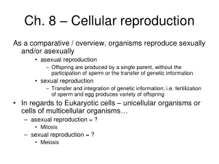
In sexual reproduction • Fertilization of sperm and egg produces offspring • In asexual reproduction • Offspring are produced by a single parent, without the participation of sperm and egg
LM 340 CONNECTIONS BETWEEN CELL DIVISION AND REPRODUCTION • 8.1 Like begets like, more or less • Some organisms reproduce asexually • And their offspring are genetic copies of the parent and of each other Figure 8.1A
Other organisms reproduce sexually • Creating a variety of offspring Figure 8.1B
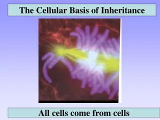
8.2 Cells arise only from preexisting cells • Cell division is at the heart of the reproduction of cells and organisms • Because cells come only from preexisting cells
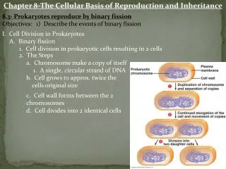
Prokaryotic chromosomes Colorized TEM 32,500 • 8.3 Prokaryotes reproduce by binary fission • Prokaryotic cells • Reproduce asexually by cell division Figure 8.3B
Duplication of chromosomeand separation of copies 1 Continued elongation of thecell and movement of copies 2 Division intotwo daughter cells 3 • As the cell replicates its single chromosome, the copies move apart • And the growing membrane then divides the cells Plasmamembrane Prokaryoticchromosome Cell wall Figure 8.3A
THE EUKARYOTIC CELL CYCLE AND MITOSIS • 8.4 The large, complex chromosomes of eukaryotes duplicate with each cell division • A eukaryotic cell has many more genes than a prokaryotic cell • And they are grouped into multiple chromosomes in the nucleus
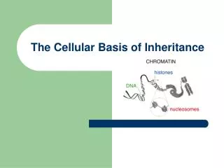
LM 600 • Individual chromosomes contain a very long DNA molecule associated with proteins • And are visible only when the cell is in theprocess of dividing • If a cell is not undergoing division • Chromosomes occur in the form of thin, loosely packed chromatin fibers Figure 8.4A
Sister chromatids Centromere TEM 36,000 • Before a cell starts dividing, the chromosomes replicate • Producing sister chromatids joined together at the centromere Figure 8.4B
Chromosomeduplication Sisterchromatids Centromere Chromosomedistributiontodaughtercells • Cell division involves the separation of sister chromatids • And results in two daughter cells, each containing a complete and identical set of chromosomes Figure 8.4C
INTERPHASE G1 S(DNA synthesis) G2 Cytokinesis Mitosis MITOTICPHASE (M) • 8.5 The cell cycle multiplies cells • The cell cycle consists of two major phases Figure 8.5
During interphase • Chromosomes duplicate and cell parts are made • During the mitotic phase • Duplicated chromosomes are evenly distributed into two daughter nuclei
8.6 Cell division is a continuum of dynamic changes • In mitosis, after the chromosomes coil up • A mitotic spindle moves them to the middle of the cell
The sister chromatids then separate • And move to opposite poles of the cell, where two nuclei form • Cytokinesis, in which the cell divides in two • Overlaps the end of mitosis
LM 250 INTERPHASE PROPHASE PROMETAPHASE Centrosomes(with centriole pairs) Fragmentsof nuclearenvelope Centrosome Early mitoticspindle Kinetochore Chromatin Nucleolus Centromere Chromosome, consistingot two sister chromatids Spindlemicrotubules Nuclearenvelope Plasmamembrane • The stages of cell division Figure 8.6 (Part 1)
TELOPHASE AND CYTOKINESIS ANAPHASE METAPHASE Nucleolusforming Cleavagefurrow Metaphaseplate Daughterchromosomes Nuclearenvelopeforming Spindle Figure 8.6 (Part 2)
Cleavagefurrow SEM 140 Cleavage furrow Contracting ring ofmicrofilaments Daughter cells • 8.7 Cytokinesis differs for plant and animal cells • In animals • Cytokinesis occurs by a constriction of the cell (cleavage) Figure 8.7A
Daughternucleus Cell plateforming Wall ofparent cell TEM 7,500 Cell wall New cell wall Vesicles containingcell wall material Cell plate Daughter cells • In plants • A membranous cell plate splits the cell in two Figure 8.7B
8.8 Anchorage, cell density, and chemical growth factors affect cell division • Most animal cells divide • Only when stimulated, and some not at all
Cells anchor todish surfaceand divide. When cells haveformed a completesingle layer, theystop dividing (density-dependent inhibition). If some cells arescraped away, theremaining cells divideto fill the dish with asingle layer and thenstop (density-dependentinhibition). • In laboratory cultures • Most normal cells divide only when attached to a surface • They continue dividing • Until they touch one another Figure 8.8A
After forming asingle layer,cells havestopped dividing. Providing anadditional supply ofgrowth factorsstimulatesfurther cell division. • Growth factors • Are proteins secreted by cells that stimulate other cells to divide Figure 8.8B
8.9 Growth factors signal the cell cycle control system • A set of proteins within the cell • Controls the cell cycle
G1 checkpoint G0 Controlsystem S G1 G2 M Mcheckpoint G2 checkpoint • Signals affecting critical checkpoints in the cell cycle • Determine whether a cell will go through the complete cycle and divide Figure 8.9A
Growth factor Plasma membrane Relayproteins Receptorprotein G1 checkpoint Signaltransductionpathway Controlsystem S G1 M G2 • The binding of growth factors to specific receptors on the plasma membrane • Is usually necessary for cell division. Figure 8.9B
CONNECTION • 8.10 Growing out of control, cancer cells produces malignant tumors • Cancer cells • divide excessively to form masses called tumors
Lymphvessels Tumor Bloodvessel Glandulartissue Cancer cells invadeneighboring tissue. Cancer cells spread throughlymph and blood vessels toother parts of the body. A tumor grows from asingle cancer cell. • Malignant tumors • Can invade other tissues Figure 8.10
Radiation and chemotherapy • Are effective as cancer treatments because they interfere with cell division
LM 500 • 8.11 Review of the functions of mitosis: Growth, cell replacement, and asexual reproduction • When the cell cycle operates normally, mitotic cell division functions in • Growth Figure 8.11A
LM 700 • Replacement of damaged or lost cells Figure 8.11B
LM 10 • Asexual reproduction Figure 8.11C
MEIOSIS AND CROSSING OVER • 8.12 Chromosomes are matched in homologous pairs • The somatic (body) cells of each species • Contain a specific number of chromosomes • For example human cells have 46 • Making up 23 pairs of homologous chromosomes
Chromosomes Centromere Sister chromatids • The chromosomes of a homologous pair • Carry genes for the same characteristics at the same place, or locus Figure 8.12
8.13 Gametes have a single set of chromosomes • Cells with two sets of chromosomes • Are said to be diploid • Gametes, eggs and sperm, are haploid • With a single set of chromosomes
Haploid gametes (n = 23) n Egg cell n Sperm cell Meiosis Fertilization Diploidzygote(2n = 46) 2n Multicellulardiploid adults(2n = 46) Mitosis and development • Sexual life cycles • Involve the alternation ofhaploid and diploid stages Figure 8.13
8.14 Meiosis reduces the chromosome number from diploid to haploid • Meiosis, like mitosis • Is preceded by chromosome duplication • But in meiosis • The cell divides twice to form four daughter cells
The first division, meiosis I • Starts with synapsis, the pairing of homologous chromosomes • In crossing over • Homologous chromosomes exchange corresponding segments • Meiosis I separates each homologous pair • And produce two daughter cells, each with one set of chromosomes
Meiosis II is essentially the same as mitosis • The sister chromatids of each chromosome separate • The result is a total of four haploid cells
MEIOSIS I: Homologous chromosomes separate INTERPHASE PROPHASE I METAPHASE I ANAPHASE I Sister chromatids remain attached Microtubulesattached to kinetochore Centrosomes (with centriole pairs) Metaphaseplate Sites of crossing over Spindle Centromere(with kinetochore) Homologouschromosomes separate Sisterchromatids Nuclearenvelope Tetrad Chromatin • The stages of meiosis Figure 8.14 (Part 1)
MEIOSIS II: Sister chromatids separate TELOPHASE IAND CYTOKINESIS TELOPHASE IIAND CYTOKINESIS PROPHASE II METAPHASE II ANAPHASE II Cleavagefurrow Sister chromatidsseparate Haploid daughter cellsforming Figure 8.14 (Part 2)
Mitosis Meiosis Parent cell(before chromosome replication) Meiosis i Prophase I Prophase Tetrad formedby synapsis ofhomologouschromosomes Chromosome replication Chromosome replication Duplicated chromosome(two sister chromatids) 2n = 4 Chromosomes align at themetaphase plate Tetradsalign at themetaphase plate Metaphase I Metaphase Anaphase ITelophase I Homologous chromosomesseparate duringanaphase I;sister chromatidsremain together Sister chromatidsseparate during anaphase AnaphaseTelophase Haploidn = 2 Daughtercells of meiosis I No furtherchromosomalreplication; sisterchromatids separateduringanaphase II Meiosis ii 2n 2n Daughter cellsof mitosis n n n n Daughter cells of meiosis II • 8.15 Review: A comparison of mitosis and meiosis Figure 8.15
8.16 Independent orientation of chromosomes in meiosis and random fertilization lead to varied offspring • Each chromosome of a homologous pair • Differs at many points from the other member of the pair
Possibility 1 Possibility 2 Two equally probablearrangements of chromosomes at metaphase I Metaphase II Gametes Combination 2 Combination 4 Combination 1 Combination 3 • Random arrangements of chromosome pairs at metaphase I of meiosis • Lead to many different combinations of chromosomes in eggs and sperm Figure 8.16
Random fertilization of eggs by sperm • Greatly increases this variation
White coat (C); pink eyes (e) Brown coat (C); black eyes (E) Eye-colorgenes Coat-colorgenes Brown Black C E C E C E Meiosis c e c e Pink White c e Tetrad in parent cell(homologous pair ofduplicated chromosomes) Chromosomes of the four gametes • 8.17 Homologous chromosomes carry different versions of genes • The differences between homologous chromosomes • Are based on the fact that they can bear different versions of a gene at corresponding loci Figure 8.17B Figure 8.17A
TEM 2,200 Chiasma Tetrad Centromere • 8.18 Crossing over further increases genetic variability • Genetic recombination • Which results from crossing over during prophase I of meiosis, increases variation still further Figure 8.18A
Coat-colorgenes Eye-colorgenes Breakage of homologous chromatids 1 Joining of homologous chromatids 2 Separation of homologous chromosomes at anaphase I 3 Separation of chromatids at anaphase II and completion of meiosis 4 • How crossing over leads to genetic variation E C Tetrad (homologous pair of chromosomes in synapsis) e c E C e c E C Chiasma e c E C e C E c e c E C Parental type of chromosome e C Recombinant chromosome E c Recombinant chromosome e c Parental type of chromosome Gametes of four genetic types Figure 8.18B
ALTERATIONS OF CHROMOSOME NUMBER AND STRUCTURE • 8.19 A karyotype is a photographic inventory of an individual’s chromosomes • A karyotype • Is an ordered arrangement of a cell’s chromosomes