Download

1 / 26
260 likes | 344 Views
Understand cell division, binary fission, chromosomes, cell cycle phases, and key factors influencing cell division in this comprehensive module.

E N D
CHAPTER 8The Cellular Basis of Reproduction and Inheritance Modules 8.1 – 8.11
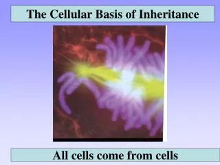
8.2 Cells arise only from preexisting cells The Cell Theory • 1. All living organisms are composed of one or more cells. (1958 Virchow) • 2. Cells are the most basic unit for function and structure of all organisms. • 3. All cells come from cells that already exist through cell division. Cellular division has several functions: • Cell division allows an embryo to develop into an adult • Cell division heals wounds when inflicted • It also ensures the continuity of life from one generation to the next
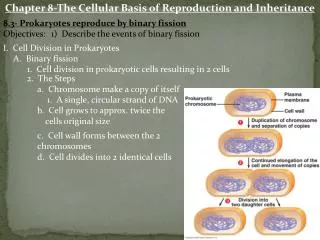
8.3 Prokaryotes reproduce by binary fission • Prokaryotic cells divide asexually • These cells possess a single chromosome, containing genes. There may be additional plasmids in bacteria. • The chromosome is replicated • The cell then divides into two cells, a process called binary fission Prokaryotic chromosomes Figure 8.3B
Plasmamembrane Prokaryoticchromosome Cell wall • Binary fission of a prokaryotic cell Duplication of chromosomeand separation of copies Continued growth of the cell and movement of copies Division intotwo cells Figure 8.3A
eukaryotic vs prokaryotic • A eukaryotic cell has many more genes than a prokaryotic cell. (A bacteria may have 3000 genes while a human may have 50,000 to 100,000) • The genes are grouped into multiple chromosomes, found in the nucleus. • To accomplish cell division, the chromosomes must go through temporary alterations in their structure. Figure 8.4A
Individual chromosomes are only visibleduring cell division when they become tightly coiled. • Chromosomes are packaged as chromatin. Chromatin is DNA and proteins. • What is the adaptive advantage of becoming tightly coiled when the cell divides? • Chromosomes are made of a very long DNA molecule with thousands of genes. The DNA in one human cell is approximately 2 meters long.
Sister chromatids • Before a cell starts dividing, the chromosomes are duplicated. This must occur so that each daughter cell gets the same amount of DNA as the cell it came from. Centromere • This process produces 2 sister chromatids which make up one chromosome. Figure 8.4B
Chromosomeduplication • Two daughter cells are produced • Each has a complete and identical set of chromosomes • When the cell divides, the sister chromatids separate Sister chromatids Centromere Chromosomedistributiontodaughtercells Figure 8.4C
8.5 The cell cycle multiplies cells • The cell cycle consists of two major phases: • Interphase, where chromosomes duplicate and cell parts are made • The mitotic phase, when cell division occurs Figure 8.5
8.6 Cell division is a continuum of dynamic changes • Eukaryotic cell division consists of two stages: • Mitosis • Cytokinesis • Let’s take a closer look.
INTERPHASE PROPHASE Centrosomes(with centriole pairs) Early mitoticspindle Centrosome Fragmentsof nuclearenvelope Kinetochore Chromatin Centrosome Spindlemicrotubules Nucleolus Nuclearenvelope Plasmamembrane Chromosome,consisting of twosister chromatids Figure 8.6
METAPHASE ANAPHASE TELOPHASE AND CYTOKINESIS Cleavagefurrow Nucleolusforming Metaphaseplate Nuclearenvelopeforming Spindle Daughterchromosomes Figure 8.6 (continued)
8.7 Cytokinesis differs for plant and animal cells • In animals, cytokinesis occurs by cleavage • This process pinches the cell apart Cleavagefurrow Cleavagefurrow Contracting ring ofmicrofilaments Figure 8.7A Daughter cells
Cell plateforming Wall ofparent cell Daughternucleus • In plants, a membranous cell plate splits the cell in two Cell wall New cell wall Vesicles containingcell wall material Cell plate Daughtercells Figure 8.7B
8.8 Anchorage, cell density, and chemical growth factors affect cell division • Most animal cells divide only when stimulated, and others not at all • In laboratory cultures, most normal cells divide only when attached to a surface • They are anchorage dependent.
This is called density-dependent inhibition • Cells continue dividing until they touch one another Cells anchor to dish surface and divide. When cells have formed a complete single layer, they stop dividing (density-dependent inhibition). If some cells are scraped away, the remaining cells divide to fill the dish with a single layer and then stop (density-dependent inhibition). Figure 8.8A
Growth factors are proteins secreted by cells that stimulate other cells to divide After forming a single layer, cells have stopped dividing. Providing an additional supply of growth factors stimulates further cell division. Figure 8.8B
8.9 Growth factors signal the cell cycle control system • Proteins within the cell control the cell cycle • Signals affecting critical checkpoints determine whether the cell will go through a complete cycle and divide G1 checkpoint Controlsystem M checkpoint Figure 8.9A G2 checkpoint
The binding of growth factors to specific receptors on the plasma membrane is usually necessary for cell division Growth factor Plasma membrane Relayproteins G1 checkpoint Receptor protein Signal transduction pathway Cell cyclecontrolsystem Figure 8.8B
8.10 Connection: Growing out of control, cancer cells produce malignant tumors • Cancer cells have abnormal cell cycles • They divide excessively and can form abnormal masses called tumors • Radiation and chemotherapy are effective as cancer treatments because they interfere with cell division
Malignant tumors can invade other tissues and may kill the organism Lymphvessels Tumor Glandulartissue Metastasis 1 A tumor grows from a single cancer cell. 2 Cancer cells invade neighboring tissue. 3 Cancer cells spread through lymph and blood vessels to other parts of the body. Figure 8.10
8.11 Review of the functions of mitosis: Growth, cell replacement, and asexual reproduction • When the cell cycle operates normally, mitotic cell division functions in: • Growth (seen here in an onion root) Figure 8.11A
Deadcells • Cell replacement (seen here in skin) Epidermis, the outer layer of the skin Dividingcells Dermis Figure 8.11B
Asexual reproduction (seen here in a hydra) Figure 8.11C