Download
1 / 27
270 likes | 422 Views
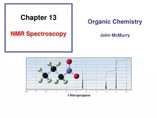
13 C NMR Spectroscopy. 1 H and 13 C NMR compared:. both give us information about the number of chemically nonequivalent nuclei (nonequivalent hydrogens or nonequivalent carbons) both give us information about the environment of the nuclei (hybridization state, attached atoms, etc.)

E N D
1H and 13C NMR compared: both give us information about the number of chemically nonequivalent nuclei (nonequivalent hydrogens or nonequivalent carbons) both give us information about the environment of the nuclei (hybridization state, attached atoms, etc.) it is convenient to use FT-NMR techniques for 1H; it is standard practice for 13C NMR
1H and 13C NMR compared: 13C requires FT-NMR because the signal for a carbon atom is 10-4 times weaker than the signal for a hydrogen atom a signal for a 13C nucleus is only about 1% as intense as that for 1H because of the magnetic properties of the nuclei, and at the "natural abundance" level only 1.1% of all the C atoms in a sample are 13C (most are 12C)
1H and 13C NMR compared: 13C signals are spread over a much wider range than 1H signals making it easier to identify and count individual nuclei Figure 1 (a) shows the 1H NMR spectrum of 1-chloropentane; Figure 1 (b) shows the 13C spectrum. It is much easier to identify the compound as 1-chloropentane by its 13C spectrum than by its 1H spectrum.
10.0 9.0 8.0 7.0 6.0 5.0 4.0 3.0 2.0 1.0 0 1H Figure 1 (a) CH3 ClCH2 ClCH2CH2CH2CH2CH3 Chemical shift (d, ppm)
200 180 160 140 120 100 80 60 40 20 0 13C Figure 1 (b) a separate, distinct peak appears for each of the 5 carbons ClCH2CH2CH2CH2CH3 CDCl3 Chemical shift (d, ppm)
13C Chemical Shifts are measured in ppm (d)from the carbons of TMS
13C Chemical shifts are most affected by: • hybridization state of carbon • electronegativity of groups attached to carbon
Examples (chemical shifts in ppm from TMS) sp3 hybridized carbon is more shielded than sp2 23 138
OH O Examples (chemical shifts in ppm from TMS) sp3 hybridized carbon is more shielded than sp2 61 202
OH Examples (chemical shifts in ppm from TMS) an electronegative atom deshields the carbon to which it is attached 23 61
O Examples (chemical shifts in ppm from TMS) an electronegative atom deshields the carbon to which it is attached 138 202
Table 1 Type of carbon Chemical shift (d),ppm RCH3 0-35 R2CH2 15-40 R3CH 25-50 R4C 30-40
RC CR R2C CR2 Table 2 Type of carbon Chemical shift (d),ppm Type of carbon Chemical shift (d),ppm RCH3 0-35 65-90 R2CH2 15-40 100-150 R3CH 25-50 110-175 R4C 30-40
Table 3 Type of carbon Chemical shift (d),ppm RCH2Br 20-40 RCH2Cl 25-50 35-50 RCH2NH2 50-65 RCH2OH RCH2OR 50-65
O O Table 4 Type of carbon Chemical shift (d),ppm Type of carbon Chemical shift (d),ppm RCH2Br 20-40 RCOR 160-185 RCH2Cl 25-50 35-50 RCH2NH2 50-65 RCH2OH RCR 190-220 RCH2OR 50-65
13C NMR and Peak Intensities Pulse-FT NMR distorts intensities of signals. Therefore, peak heights and areas can be deceptive.
CH3 OH 200 180 160 140 120 100 80 60 40 20 0 Figure 2 7 carbons give 7 signals, but intensities are not equal Chemical shift (d, ppm)
Peaks in a 13C NMR spectrum are typicallysinglets 13C—13C splitting is not seen because the probability of two 13C nuclei being in the same molecule is very small. 13C—1H splitting is not seen because spectrum is measured under conditions that suppress this splitting (broadband decoupling).
Using DEPT to Count the HydrogensAttached to 13C Distortionless Enhancement of Polarization Transfer
Measuring a 13C NMR spectrum involves 1. Equilibration of the nuclei between the lower and higher spin states under the influence of a magnetic field 2. Application of a radiofrequency pulse to give an excess of nuclei in the higher spin state 3. Acquisition of free-induction decay data during the time interval in which the equilibrium distribution of nuclear spins is restored 4. Mathematical manipulation (Fourier transform) of the data to plot a spectrum
Measuring a 13C NMR spectrum involves Steps 2 and 3 can be repeated hundreds of timesto enhance the signal-noise ratio2. Application of a radiofrequency pulse to give an excess of nuclei in the higher spin state 3. Acquisition of free-induction decay data during the time interval in which the equilibrium distribution of nuclear spins is restored
Measuring a 13C NMR spectrum involves In DEPT, a second transmitter irradiates 1H during the sequence, which affects the appearanceof the 13C spectrum. some 13C signals stay the same some 13C signals disappear some 13C signals are inverted
O CCH2CH2CH2CH3 O 200 180 160 140 120 100 80 60 40 20 0 Figure 3 (a) CH CH CH2 CH CH2 CH3 CH2 C C Chemical shift (d, ppm)
O CCH2CH2CH2CH3 200 180 160 140 120 100 80 60 40 20 0 Figure 3 (a) CH CH CH3 CH CH2 CH2 CH2 Chemical shift (d, ppm)
O CCH2CH2CH2CH3 200 180 160 140 120 100 80 60 40 20 0 Figure 3 (b) CH CH CH3 CH CH and CH3 unaffected C and C=O nulled CH2 inverted CH2 CH2 CH2 Chemical shift (d, ppm)