Analysis of DUF724 Proteins: Sequence Alignment, Phylogenetics, and Expression Patterns in Plants
This study presents a comprehensive analysis of the DUF724 protein family in plants. Supplementary Figures illustrate amino acid sequence alignments, phylogenetic trees, and gene expression patterns using data from microarray databases. Identical and similar amino acids in DUF724 domains are highlighted, and the evolutionary relationships among 18 DUF724 proteins from Arabidopsis, rice, and other species are explored. Expression data reveals distinct activity in various plant tissues, contributing to our understanding of DUF724's role in plant biology.

Analysis of DUF724 Proteins: Sequence Alignment, Phylogenetics, and Expression Patterns in Plants
E N D
Presentation Transcript
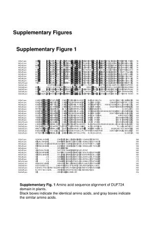
Supplementary Figures Supplementary Figure 1 Supplementary Fig. 1 Amino acid sequence alignment of DUF724 domain in plants. Black boxes indicate the identical amino acids, and gray boxes indicate the similar amino acids.
ViDuf1 91 PtDuf1 PtDuf2 100 AtDuf1 AtDuf3 90 100 97 AtDuf6 AtDuf8 AtDuf9 100 97 100 AtDuf10 100 AtDuf2 95 AtDuf5 AtDuf7 AtDuf4 OsDuf2 OsDuf3 OsDuf1 OsDuf4 100 OsDuf5 Supplementary Figure 2 Supplementary Fig. 2 Phylogenetic analysis of DUF724 proteins in plants. Phylogenetic tree was constructed by deduced full-length amino acid sequences of 18 DUF724 proteins in Arabidopsis, rice, poplar and Vitis vinifera.
AtDuf10 AtDuf9 AtDuf8 AtDuf3 AtDuf6 AtDuf1AtDuf2 AtDuf7 AtDuf5AtDuf4 callus cell suspension seeding cotyledons hypocotyl radicle inflorescence flower petal sepal stamen pedicel silique seed stem node shoot apex cauline leaf rosette juvenile leaf adult leaf petiole senescent leaf hypocotyle roots root tip elongation zone root hair zone stele germinated seed seedling young rosetted developed rosette bolting young flower developed flower flowers and Siliques mature siliques Color scale >=95% >=90%<95% >=80%<85% AtDuf10 AtDuf9 AtDuf8 AtDuf3 AtDuf6 AtDuf1AtDuf2 AtDuf7 AtDuf5AtDuf4 >=70%<75% >=60%<65% >=50%<55% >=40%<45% >=30%<35% >=20%<25% >=10%<15% >=0%<5% Supplementary Figure 3 Supplementary Fig. 3 The expression patterns of the AtDUF724 genes. The figure was prepared using the data obtained from the public microarray databases through Genevestigator, showing the expression patterns of AtDuf genes in different tissues.
Myc blot HA blot 4 5 6 MW 3 1 2 MW 95 95 * * 72 72 55 55 * 43 * 43 * * Supplementary Figure 4 (A) (B) Supplementary Fig. 4 Expression of GAL4 AD-AtDuf and GAL4 BD-AtDuf fusion proteins in yeast two hybrid system The protein lysates harvested from yeasts transformed with different constructs were assayed by western blotting with antibody against the HA tag (a) and Myc tag (b). Lane 1, pGADT7-AtDuf5; lane 2, pGADT7-AtDuf7; lane 3, pGADT7-AtDuf3; lane 4, pGBKT7-AtDuf5; lane 5, pGBKT7-AtDuf7; lane 6, pGBKT7-AtDuf3 and MW, molecular weight. Stars indicate the position of individual GAL4 fusion proteins.