Download
1 / 33
410 likes | 844 Views
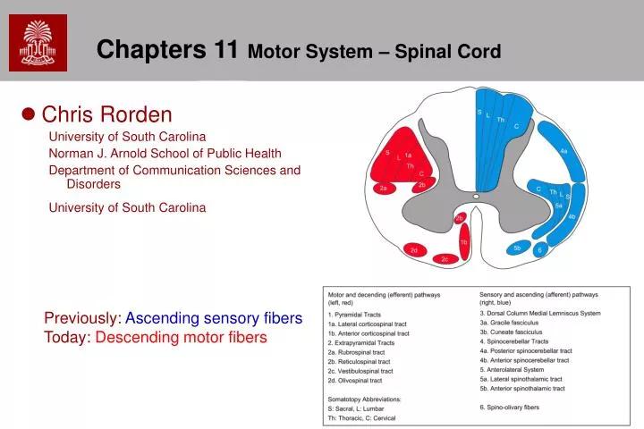
Chapters 11 Motor System – Spinal Cord. Chris Rorden University of South Carolina Norman J. Arnold School of Public Health Department of Communication Sciences and Disorders University of South Carolina. Previously: Ascending sensory fibers Today: Descending motor fibers.

E N D
Chapters 11 Motor System – Spinal Cord • Chris Rorden University of South Carolina Norman J. Arnold School of Public Health Department of Communication Sciences and Disorders University of South Carolina Previously: Ascending sensory fibers Today: Descending motor fibers
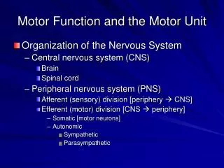
Six Neuraxial* Levels • Spinal Cord • Brainstem • Cerebellum • Diencephalon • Basal Ganglia • Cerebral Cortex *Neuraxial = Brain and Spinal Cord Axis
Functional Levels • Spinal Level • Simple Reflexes • Regulation of Higher Skilled or Patterned Movements • Upper Levels • Initiation, Inhibition or Facilitation of Motor Functions • Voluntary Motor Movements
Spinal Cord • 43.5cm long, 1cm diameter • Five Spinal Segments and Spinal Nerve Groups • Cervical (8) • Thoracic (12) • Lumbar (5) • Sacral (5 fused vertebrae), "Holy Bone" • Coccygeal (3-5 fused vertebrae) ‘tailbone’, (coccyx = cuckoo's beak)
Spine and Pelvis • Spine can rotate with respect to pelvis
Spinal Nerves • There are a total of 31 bilaterally-paired spinal nerves • 8 cervical nerves (C1-C8) • 12 thoracic nerves (T1-T12) • 5 lumbar nerves (L1-L5) • 5 sacral nerves (S1-S5) • 1 coccygeal nerve (Co, skin of lower back) • C1 to C7 exit vertebral canal above the respective cervical vertebra (e.g. C1 exits above the first cervical vertebra). • All the other spinal nerves (C8, T*, L*, S*,Co) leave below their corresponding vertebra.
Spinal Cord and Vertebrae Yellow=CSF • Vertebral Column Longer Than Spinal Cord • Conus Medullaris • End of Spinal Cord at L2 • Cauda Equina (Horse’s Tail – nerve roots) • Stretched nerve root fibers from L3 to S5 • Filum Terminale - fibrous tissue • Stretched Spinal Cord Remnant Attached to Coccyx • Cauda equina • Contains Lumbosacral Cistern • Fluid Filled Space for Spinal Puncture • Spinal cord stops growth during infancy, spine grows through adolescence.
Lumbar Nerves L1 Conus medullaris L2 Cauda equina (horses tail) saddle area, sphincters, parasympathetic Bladder/bowel L3 L4 L5 Filum terminale
Sacral Nerves S1 S2 S3 S4 S5 Co • Filum terminale • coccygeal ligament. • connective tissue (pia mater) • From medullary cone to the termination of the vertebral canal.
Meninges • "The meninges PAD the brain and spinal column." -- Pia; Arachnoid; Dura. • Dentate Ligaments • The pia mater has 21 pairs of denticulate ligaments which attach it to the arachnoid and dura maters. • provide stability for the spinal cord against motion within the vertebral column.
Spinal Cord • Internal Structure • White Matter – outer parts of the cord • Gray Matter Horns and Commissures – the internal sections • Varies in Shape With Level of Spinal Segment • Dorsal Root and Root Ganglia • Ventral Root
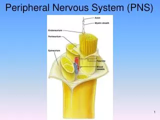
Spinal Nerves • Exit Vertebra Through Intervertebral Foramina • Dorsal and Ventral Rami Form Spinal Nerve • Dorsal Roots - Sensory Information • Ventral Roots - Motor Information • Except between T-2 and T-11, Ventral Roots Form Plexi to Serve Groups of Muscles • A nerve plexus is a network of intersecting nerves. They combine sets of spinal nerves that serve the same area of the body into one large grouped nerve.
Cross Section • Gray matter of the Spinal Cord • Dorsal Horn • Ventral Horn
Cross sections L4 T2 White Matter (tracts) S3 Gray Matter (interneurons)
Motor Units • Lower Motor Neuron • Lower motor neurons (LMNs) are the motor neurons bring the nerve impulses from the upper motor neurons out to the muscles. • Path for Efferent Impulses • Final Common Pathway (to Muscles) • Four Components • Motor Cell body • Efferent Fiber • Motor End Plate - Myoneural-Neuromuscular Junction • Muscle Fibers Innervated by Axon
Tracts of Spinal Cord • Neural impulses are carried through white matter • Three Major Bundles • Dorsal Column: Primarily Ascending Fibers • Lateral: Ascending and Descending Fibers • Anterior (aka Ventral): Ascending and Descending Fibers
Descending Tracts • Pyramidal (aka Corticospinal) Tracts • From cortex – Betz Cells (large pyramidal cells) in precentral gyrus. • Through Internal Capsule, Pes Pedunculi, Pontine Nuclei, Pyramidal Decussation (medulla): 90% decussate, Spinal Cord • Extrapyramidal Tract • not directly from motor or premotor cortex • Autonomic Pathways • pathways from thalamus to spinal cord and brainstem – regulates motor functions of the sympathetic and parasympathetic systems (inspiration, vomiting, and coughing reflexes)
Upper Motor Neurons • Upper motor neurons: motor neurons that are NOT directly responsible for stimulating the target muscle
Upper Motor Neurons • Upper motor neurons Tracts • Cortico-spinal: motor cortex to spinal nerve roots – fine voluntary movements • Corticobulbar: Cortex to pons and medulla – involuntary maintenance of posture • Tectospinal – Superior Colliculus to lower motor neurons. Involuntary correction of head to visual stimuli • Rubrospinal: red nucleus to LMN • Vestibulospinal: vestibular nuclei- responsible for adjusting posture to maintain balance. • Reticulospinal: reticular formation - balance
Corticospinal fibers LCT • Lateral Corticospinal Tract • Control of Skeletal Muscle (Fingers, Toes, Forearm) • Skilled Manipulations • 90% Decussate and Form Alpha Fibers in Ventral Horn • Anterior Corticospinal Tract (AKA ventral corticospinal tract) • 8-10% Fibers That Did Not Cross Midline • Cross at Spinal Horn • Control Axial and Girdle Muscles – responsible for moving head axial movement of head and trunk ACT
Descending Tracts • Tectospinal Tract • Response to Visual Stimulation • Superior Colliculus to Cervical Spinal Cord • Rubrospinal Tract • Regulation of Muscle Tone Against Gravity • Red N. To Motor Nerve Cells in Ventral Horn • Vestibulospinal Tract • Reflexive Adjustment of Body and Limbs • Vestibular N. To Spinal Cord • Reticular Descending Tract • Alteration of Muscle Tone
Descending Autonomic Tracts • Hypothalamus: • Projects to Brainstem and Spinal Visceral Nuclei • Regulate Autonomic Function of Sympathetic and Parasympathetic Systems
Ascending (Sensory) Tracts • Fasciculus Gracilis • Fasciculus Cuneatus • Anterior Spinothalamic Tract • Lateral Spinothalamic Tract • Ventral Spinocerebellar Tract • Dorsal Spinocerebellar Tract • Cuneocerebellar Tract • Spinotectal Tract • Spinoreticular Tract
Types of Motor Nerve Cells • Anterior Motor Neurons – exit at the ventral horns • Alpha and Y (Gamma) Motor Nerve Cells • Lower Motor Neurons (Below 2nd Level in Neuronal Pathway) • Interneurons • Association Cells Connecting Sensory and Motor Neuron Pools • Often Part of Reflexive Action
Motor Neurons • Alpha Neurons • Major Motor Neurons • Small • Responsible for Voluntary and Reflexive Movements of Head, Trunk and Extremities • One Fiber Can Innervate 200 Muscles fibers • Y-Motor Neurons • Smaller and Fewer • Controlled by Reticular and Vestibular Systems
Interneurons • 30 Times More Than Motor Neurons • Filter of Sensory and Motor Function • Function As Inhibitory Cells and Association Cells
Motor functions of the spinal cord • Reflexive Motor Response • Stereo-Typical (Rote) Response to Stimulus • Involves Muscle Spindles, Afferent Fibers, Alpha Motor Neurons, Efferent Fibers and Muscles • Independent of Voluntary Control • Upper Centers Become Involved to Smooth Reaction and Return body to homeostasis
Muscle Receptors • Two Types of Receptors • Muscle Spindle • Sensor inside muscle • Detects and Maintains Muscle Tension • Golgi Tendon Organs • Sensor on tendon • Monitors Degree of Muscle Tension During Contraction • Prevents Too Much Tension
Spinal Reflexes • Stretch Reflex i.e. knee reflex • Tap Patella causing tendon change (y motor neuron) • Muscle spindles stimulate alpha motor neuron response, and muscle contracts • Occurs at the L3 level • Withdrawal (Flexor) Reflex i.e. Touching Hot Stimulus • Protective Response to pain • Flexion of leg or arm • Stimulus, receptor, substantia gelatinosa, interneurons and alpha neuron response
Spinal Reflexes • Crossed (Intrasegmental) Extensor Reflex • Protective response • Involves both sides of the body • As one arm is withdrawn, the other arm is extended • Multisynaptic because it involves opposite body parts • An example of this is when a person steps on a nail, the leg that is stepping on the nail pulls away, while the other leg takes the weight of the whole body.
Neurotransmitters • Spinal Cord (excitatory) • Epinephrine • Norepinephrine • Serotonin • PNS • Acetylcholine
Clinical Considerations • Many Sources of Lesions • Trauma • Tumors or Infections • Degenerative Conditions • Compare the Function of One Side to the Other • Hyper Quality of Movement (Spastic) (Upper Motor Neuron Problems) • Hypo Quality of Movement (Spinal or Spinal Nerve Level - Lower Motor Neuron) Causing Flaccid Paralysis • Absent Reflexes and Atrophy or Muscle Wasting
Common Spinal Syndromes • Complete Spinal Transection • Dislocations, tumor, myelitis • Function lost below the lesion • After a period of time, reflexes may become spastic in nature • Brown-Sequard Syndrome (cord tumor, trauma, ischemia) • Lesion on ipsilateral half of body, ipsilateral sensory loss, contralateral pain and temperature sensation loss • Syringomyelia • Developmental condition: cyst formation within spinal cord with loss of sensation and muscle control – usually starts between ages 25-40