Download
1 / 55
740 likes | 1.58k Views
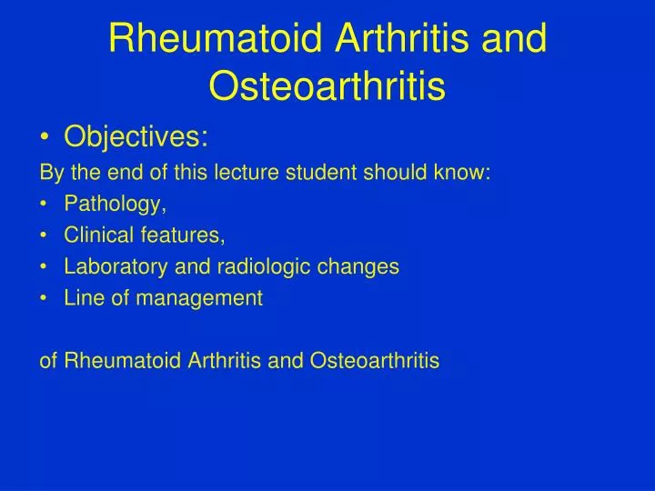
Rheumatoid Arthritis and Osteoarthritis. Objectives: By the end of this lecture student should know: Pathology, Clinical features, Laboratory and radiologic changes Line of management of Rheumatoid Arthritis and Osteoarthritis. Rheumatoid Arthritis Systemic chronic inflammatory disease

E N D
Rheumatoid Arthritis and Osteoarthritis • Objectives: By the end of this lecture student should know: • Pathology, • Clinical features, • Laboratory and radiologic changes • Line of management of Rheumatoid Arthritis and Osteoarthritis
Rheumatoid Arthritis Systemic chronic inflammatory disease Mainly affects synovial joints Variable expression Prevalence about 3% Worldwide distribution Female:male ratio 3:1 Peak age of onset: 25-50 years
Rheumatoid Arthritis • Unknown etiology • Genetics • Environmental • Possible infectious component • Autoimmune disorder
THE PATHOLOGY OF RA • Synovitis Joints Tendon sheaths Bursae • Nodules • Vasculitis
RA Is Characterised by Synovitis and Joint Destruction NORMAL RA Inflamed synovial membrane Synovial membrane • Major cell types: • T lymphocytes • macrophages Pannus Cartilage • Minor cell types: • fibroblasts • plasma cells • endothelium • dendritic cells Synovial fluid • Major cell type: • neutrophils Capsule Cartilage thinning Adapted from Feldmann M, et al. Annu Rev Immunol. 1996;14:397-440.
Numerous Cellular Interactions Drive the RA Process Immune complexes Bacterial products IL-1, TNF-, etc Rheumatoid factors B cell IL-1 Soluble factors and direct cell–cell contact T cell HLA -DR Antigen- presenting cells Macrophage B cell or macrophage IL-1 and TNF- Synoviocytes Chondrocytes Pannus Articular cartilage Production of collagenase and otherneutral proteases Arend W. Semin Arthritis Rheum. 2001;30(suppl 2):1-6.
IL-1 and TNF- Have a Number of Overlapping Proinflammatory Effects Proinflammatory effects of IL-1 Proinflammatory effects of TNF- COX-2PGE2NOAdhesion moleculesChemokinesCollagenasesIL-6 TNF-Osteoclast activation Angiogenic factors IL-1 cell death COX-2 = cyclo-oxygenase type 2; PGE2 = prostaglandin-E2; NO = nitric oxide
IL-1 Plays a Pivotal Role in the Inflammatory and Destructive Processes of RA IL-1 Activates monocytes/macrophages Induces fibroblast proliferation Activates chondrocytes Activates osteoclasts Inflammation Synovial pannus formation Cartilage breakdown Bone resorption
Signs and Symptoms • Joint inflammation • Tender, warm swollen joints • Symmetrical pattern • Pain and stiffness • Symptoms in other parts of the body • Nodules • Anemia • Fatigue, occasional fever, malaise
JOINT INVOLVEMENT ON PRESENTATION OF RA Polyarticular 75% Monoarticular 25% Small joints Knee 50% of hands and feet 60% Large joints 30% Shoulder } Wrist } Large and Hip } 50% Small joints 10% Ankle } Elbow }
Articular features seen in the Rheumatoid Hand WRIST:PIPs: Synovitis Synovitis Prominent ulnar styloid Fixed flexion or extension Subluxation and collapse of deformities carpus (Swan neck or boutonniere Radial deviation deformity) MCPs: THUMBS: Synovitis Synovitis Ulnar deviation ‘Z’ deformity Subluxation
Extra-articular manifestations • General • fever, lymphadenopathy, weight loss, fatigue • Dermatologic • palmar erythema, nodules, vasculitis • Ocular • episcleritis/scleritis, scleromalacia perforans, choroid and retinal nodules
Extra-articular manifestations • Cardiac • pericarditis, myocarditis, coronary vasculitis, nodules on valves • Neuromuscular • entrapment neuropathy, peripheral neuropathy, mononeuritis multiplex • Hematologic • Felty’s syndrome, large granular lymphocyte syndrome, lymphomas
Extra-articular manifestations • Pulmonary • pleuritis, nodules, interstitial lung disease, bronchiolitis obliterans, arteritis, effusions • Others • Sjogren’s syndrome, amyloidosis
Investigations: • Hematology : CBC , ESR • Biochemistry : LFT , Renal profile • Serology : RF , Anti-CCP • Radiography : Joints , Spines ,Chest
The 2010 ACR / EULAR classification criteria for rheumatoid arthritis Target population (Who should be tested?): Patients who 1) have at least1 joint with definite clinical synovitis (swelling) 2) with the synovitis not better explained by another disease Add A–D; a score of 6/10 is needed to classify patient as having definite RA A.Joint involvement 1 large joint. 0 2-10 large joints 1 1-3 small joints (with or without involvement of large joints) 2 4-10 small joints (with or without involvement of large joints) 3 3-10 joints (at least 1 small joint) 5 B. Serology (at least 1 test result is needed for classification) Negative RF and negative ACPA 0 Low-positive RF or low-positive ACPA 2 High-positive RF or high-positive ACPA 3 C. Acute-phase reactants (1 test result is needed for classification) Normal CRP and normal ESR 0 Abnormal CRP or abnormal ESR 1 D. Duration of symptoms 6 weeks 0 >6 weeks 1
Treatment Goals • Relieve pain • Reduce inflammation • Prevent/slow joint damage • Improve functioning and quality of life
Treatment Approaches • Lifestyle modifications • Rest • Physical and occupational therapy • Medications • Surgery
Rationale for the Early Treatment of R.A. •Erosions develop early in the disease course •Destruction is irreversible •Disease activity is strongly associated with joint destruction later in the disease course •Early treatment can slow down radiographic progress •Disease activity must be suppressed maximally in its early stages to prevent destruction and preserve function
Drug Treatments • Nonsteroidal anti-inflammatory drugs (NSAIDs) • Disease-modifying antirheumatic drugs (DMARDs) • Biologic response modifiers • Corticosteroids
Traditional NSAIDs Aspirin Ibuprofen Ketoprofen Naproxen COX-2 Inhibitors Celecoxib Rofecoxib Nonsteroidal Anti-Inflammatory Drugs (NSAIDs)
Nonsteroidal Anti-Inflammatory Drugs (NSAIDs) • To relieve pain and inflammation • Use in combination with a DMARD • Gastrointestinal side effects
Hydroxychloroquine Sulfasalazine Methotrexate Leflunomide Gold Azathioprine Disease-Modifying Antirheumatic Drugs (DMARDs)
Disease-Modifying Antirheumatic Drugs (DMARDs) • Control symptoms • No immediate analgesic effects • Can delay progression of the disease (prevent/slow joint and cartilage damage and destruction) • Effects generally not seen until a few weeks to months
DMARDs • hydroxychloroquine • mild non-erosive disease • combinations • 200 mg bid • eye exams
DMARDs • Sulfasalazine • 1 gm bid - tid • CBC, LFTs • onset 1 - 2 months • Methotrexate • most commonly used drug • fast acting (4-6 weeks) • po, SQ - weekly • CBC, LFTs
Biologic Response Modifiers • Etanercept • Infliximab • Adalimumab • Tocilizumab
MULTIFACTORAL ETIOLOGY OF OA ● Joint instability ● Age ● Hormonal factors ● Trauma ● Altered biochemistry ● Inflammation ● Genetic predisposition ● ? Others
SYMPTOMS AND SIGNS OF OA • Pain – worse on use of joint • Stiffness – mild after immobility • Loss of movement • Pain on movement/restricted range • Tenderness (articular or periarticular) • Bony swelling • Soft tissue swelling • Joint crepitus
RADIOLOGICAL FEATURES OF OA • Narrowing of joint space • Osteophytosis • Altered bone contour • Bone sclerosis and cysts • Periarticular calcification • Soft-tissue swelling