Download
1 / 29
440 likes | 988 Views
CHAPTER 5. SIGNALING PATHWAYS IN CANCER. BY P.B.Tirupathi Pichiah 1st Year Master of Science in Animal Biotechnology. Dept of Animal Science. Bharathidasan University, Trichy. TOPICS TO BE DISCUSSED. RAS-RAF-MAP kinase pathway. Phospotidylinositol-3 kinase signaling pathway. mTOR

E N D
CHAPTER 5 SIGNALING PATHWAYS IN CANCER . BY P.B.Tirupathi Pichiah 1st Year Master of Science in Animal Biotechnology. Dept of Animal Science. Bharathidasan University, Trichy.
TOPICS TO BE DISCUSSED • RAS-RAF-MAP kinase pathway. • Phospotidylinositol-3 kinase signaling pathway. • mTOR • Translation control and growth. • Cytokine signaling • JAK / STAT pathways • Growth signaling via cell adhesion- the Wnt-β-catenin pathway. • Notch signaling • NUCLEAR PROTO-ONCOGENES AND TRANSCRIPTION FACTORS.
RAS-RAF-MAP kinase pathway. • RAF is the first member of three-kinase modular sequence, with generic designation MAPKKK,MAPKK,MAPK. • MAPK-Mitogen Activated Protein Kinase. • More than 6 distinct pathway follow this general pattern in mammalian cells including JUN kinase/stress activated kinase (SAPK) • The mitogenic pathway involves RAF, MAPK/ERK kinase (MEK1 and 2) and extracellular receptor kinase (ERK1 and 2). • Sequential kinase cascades may function to amplify the input signal. • Linking of some scaffold proteins such as MP1 (MEK Partner) and JIP1 (JUN N-terminal kinase interacting protein) prevents signal amplification.
RAS-RAF-MAP kinase pathway. • Prevents undesirable spread of signaling across multiple pathway. • The end result of MAPK sequence is relocation of active ERK to the nucleus , and phosphorylation of regulated transcription factors of the ETS family. • Ultimately this lead to enhanced expression of other key transcription factors such as c-MYC, which is key to cell cycle control.
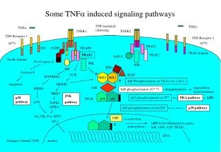
LIGAND RTK (e.g. IGF) PIP2 DAG PLCγ PI3k p85 (SH2 domain) PI3k p110 (catalytic subunit) PDK1 IRS PIP3 P PDK1 PKC P LKB1 P PIP2 P AMPK AMPK PTEN AKT AKT P TSC2 TSC2 TSC1 TSC1 GSK3β IKK BAD PAR-4 FOXO RHEB NFкB P APOPTOSIS S6K1 S6K1 MTOR P p21K1P1 , p21CIP1 4EBP1 4EBP1 Induction of genes for cellular replication, growth and apoptosis suppression e1F4E Fig. 5.17 Lipid signaling from receptor tyrosine kinase (such as IGF-IR, HER2/Neu,VEGF-R,PDGF-R)
Phosphatidylinositol-3 kinase signaling pathway. • Phosphatidylinositol-3 kinase (PI3K) signaling pathway is as important as theRAS-MAP kinase pathway in cell survival and proliferation. • PI3K constitute lipid kinase family characterized by their ability to phosporylate inositol 3’-OH group in inositol phospholipids to generate the second messenger Phosphatidylinositol-3,4,5-triphosphate (PIP-3) • PIP-3 activates down stream kinases – AKT / PKB and S6K. • PIP-3 activates the serine / threonine kinase AKT by promoting translocation to the inner membrane where it is Phosporylated and activated by PDK1 and PDK2. • Then in turn ATK activates numerous substrate in regulation of cell survival, cell cycle progression and cellular growth, including NF-kappa B, mTOR (and thence S6K),Forkhead,BAD,GSK3β, and MDM2.
Phosphatidylinositol-3 kinase signaling pathway. • Growth factors such as insulin and IGFs can activated PI3K-AKT pathway via the IRS-2 adaptor protein recruited to activate RTK.(imp in beta cell growth regulation) • Deregulation of PI3K- CANCER due to loss of the Tumor Supressor Protein PTEN (Phosphate and tensin homolog) • Also may lead to cytoskelton deformability and motility. • AKT is hyper activated in Human cancer. • AKT activation is regulated by two tumor suppressors, PTEN and TSC1/TSC2 • PTEN = Breaks PIP-3 to PIP2 = inhibit PI3K upstream of AKT.
Phosphatidylinositol-3 kinase signaling pathway. • TSC1/TSC2 = Heterodimer = acts on downstream of AKT & upstream of mTOR. • PTEN & p53 tumor suppressors are among the most comonly inactivated or mutated gene in human cancer. • Inactivation of PTEN = Growth arrest through the p53 dependent cellular senesence pathway both in vitro and in vivo. • Activation of the PI3K – AKT pathway renders cancer cells resistant to apoptotic signals and promote tumor growth.
Phosphatidylinositol-3 kinase signaling pathway. • Inhibition of GSK3β is one important contributor as is phosphorylation of BAD. • Recent studies suggest that AKT may prevent apoptosis in some cases by phosphorylation of proapoptotic protein, PAR-4. • Suppressing AKT activation by PTEN cause apoptosis in cancer cells.(Goswami et al ). • Apoptosis resulting from inhibition of AKT was blocked by inhibition of other apoptosis agonist that are AKT substrate.
mTOR. • mTOR = mammalian Target Of Rapamycin protein. • downstream effector of the PI3K / AKT signaling. • mTOR is known to be involved in key cellular processes such as cell size, survival, and proliferation. (membrane trafficking, protein degradation, protein kinase C signaling, ribosome biogenesis and transcription, activates 40S ribosomal protein S6 kinase (p70s6k) and the eukaryotic initiation factor 4E-binding protein 1 ( EIF4E). • Survival signals generated by PI3K and phospholipase D target mTOR, which in turn contributes to supression of apoptotic pathways in cancer cells.
mTOR. • mTOR prevent G1 / S transition by non spesific inhibition of cell growth and also preventing CDK activation. • HAMARTOMAS :- Autosomal dominant, Germline mutation of TSC1 or TSC2, tumor development. TSC1 = hamartin and TSC2 = tuberin. ( both responsible for regulation of Cell and Organ size ) PI3 AKT mTOR S6K • ATK phosphorylates TSC2 in TSC1 / TSC2 protein complex inactivating it. While TSC1 / TSC2 has GAP activity for RHEB and activated RHEB-GTP activates mTOR. • Cell lacking TSC1 or TSC2 there is are increased levels of RHEB-GTP, which leads to activation of mTOR, leading to cell size increase and growth.
Translation control and growth • Cell proliferation needs general increase protein synthesis and a specific increase in the synthesis of replication-promoting proteins. • RAS-MAPK-and PI3K-AKT-mTOR pathways are particular. • Translation eIF Ribosomal Subunit + mRNA . eIF4 = eIF4E, eIF4G, eIF4A, eIF4B. • Polypeptide chain initiation involves the assembly of 43S initiation complex catalyze by polypeptidechain initiation factor eIF2 and the binding of eIF4E to eIF4G during the recruitment of mRNA to the ribosome. • eIF2 activity is controlled by phosphorylation of the alpha subunit done by various kinases (GCN2, and eIF2 alpha kinase 4)
Translation control and growth • Different eIF4 protein promote or inhibit translation of specific mRNAs. • mTOR – by S6K and eIF4E. • Deregulation of gene expression at the level of mRNA translation can contribute to cell transformation and the malignant phenotype. • eIF4E cooperate with c-MYC in B-cell lymphomagenesis ; in this case c-MYC could overcome growth arrest triggered by deregulated eIF4E and eIF4E could prevent c-MYC dependent apoptosis. • eIF4E over expression – head & neck, prostrate, Breast, Overian, bladder, lung cancer.
Translation control and growth • eIF4E regulates the requirement of mRNA to ribosome and thereby regulates cap-dependent protein synthesis, eIF4E also selectively enables translation of a select number of directly cancer relevent mRNAs, such as those encoding cyclin D1, c-MYC, MMP-9, and VEGF. • eIF2 and various regulatory kinase play key role in regulation of protein synthesis in respond to stresses. Phosphorylation of eFI2 reduces general protein synthesis, but increases the translation of specific mRNA that codes transcripton factors.
Cytokine Signaling. • Cytokines and growth factors activates the MAP kinase pathway resulting in the stimulation of ERK1 / 2, c-Jun N-terminal kinases, and p38 kinase, which in turn activate transcription factors like AP-1 and AP-2. • Pro-inflammatory agents such as TNF-α and IL-1 can activate the transcription factor NF-кB that in turn regulate the expression of immediately early genes involve in the immune acute phase and in inflammatory responses. • NF-кB and AP-1 are immediate-early transcriptional activators, component of the JAK / STAT pathway play an important role in the transcriptional activation of many inflammatory genes.
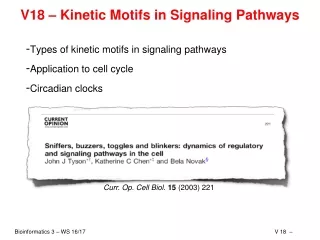
JAK / STAT pathways • JAK-STAT pathway comprises 3 families of proteins. • Janus kinases (JAK) • Signal Transducers and Activators of Transcription (STAT) • Their endogenous inhibitor SOCS family. • JAK-STAT pathway is utilized by various cytokines and growth factors. • Defective JAK-STAT-SOCS pathway may impair tumour responses to immunotherapy. • This pathway controls cell growth, cell differentiation , senescence, and apoptosis.
JAK / STAT pathways • STAT- family proteins are latent cytoplasmic transcription factor that convey signals from cytokine and growth factor receptors to the nucleus. • Binding of cytokine to cell surface receptor results in two categories of signaling.. • Activation of Cytoplasmic Tyrosine Kinase ( JAK or SRC kinase family ) • Receptor intrinsic tyrosine kinase activity (PDGF, EGF) • Tyrosine +P = activates STAT monomer = dimerize thrrough interaction of SH2 domains. (SRC homology 2) • Resultant STAT dimer translocate to STAT specific site known as gamma – activated sites (GAS) of target gene to induce transcription.
JAK / STAT pathways • STAT proteins especially STAT3 and STAT5 regulate the apoptosis resistant, angiogenesis and evade immune surveillance in tumor cells. • Epigenetic silencing of the SOCS-1 gene, an inhibitor of JAK/STAT pathway, has been described in liver cancer. • Constitutive activation of the JAK/STAT pathway is known to occur in HTLV-I-transformed T cells, Sezary’s syndrome, and v-abl or v-src transformation. • STAT3 and 5, most frequently observed in cancers (head, neck, lung, kidney,prostrate, breast, ovarian and blood)
JAK / STAT pathways • Aberrant signaling through a number of upstream pathways can result in constitutively activated STAT in tumor cells. • cellular transformation with v-src or BCR-ABL activates STAT3 and STAT5 and constitutively active STAT3 will transform immortalized fibroblast. • STAT5 can control transcription of various factors implicated in cancer including BCL-XL , PIM-1, and cyclin D1 suggesting a role both in resistant to apoptosis and replication . • JAK tyrosine kinase are activated by interleukin and other growth factors, and promote survival and proliferation of cell in multiple tissue and are constitutively active in many hematopoitic malignancies and certain carcinoma.
JAK / STAT pathways • JAK can activates both STAT and PI3K signaling. • JAK activity is essential for lymphoma invasion and metastasis, independent of its role in survival and proliferation, and independent of STAT and PI3 signaling. • Therapy targeting JAK-STAT pathway – tryphostin (AG 490), a JAK protein kinase inhibitor ,and sant 7, an IL-6 receptor antagonist.
INFα Cytokine receptor ( interferon α receptor) autophosphorylationof JAK Receptorphosphorylation INFαR Dimer Docking of STAT and their phosphorylation dimerization DNA respond element JAK- STAT PATHWAY
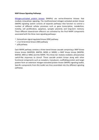
Growth signaling via cell adhesion-the Wnt-β-catenin pathway. • Cell-cell adhesiveness is decreased in cancer, enabling deregulated proliferation, migration, invasion and metastases, but in general only if cells can survive loss of cell-cell contacts. • E-caderin and interacting catenins = connects cadherin to cytoskelton. • silencing of E-caderin gene by DNA hypermethylation frequent in precancerous condition. • β-catenin also connects the E-catenin to Wingless/Wnt signaling pathway. • Specification and localization of new cells.
Growth signaling via cell adhesion-the Wnt-β-catenin pathway. • Wnt ligand binds to “Frizzled” membrane receptor and interfere with the multi-protein destruction complex, resulting in down stream activation of gene transcription by β-catenin. • multi-protein destruction complex require presence of Axin +APC (Adenomatosis polyposis coli ) acts as scaffolds to facillate phosphorylation of beta-catenin by enzyme GSK3β. P PROTEOSOME +nuclear transcription factor = expression of cMYC,cJUN,Fra,Cyclin D1 β-catenin β-catenin
Frizzled Dishevelled GPP AXIN APC AXIN APC GSK3 β-Catenin GSK3 Degradation in the proteosome β catenin TCF c-MYC, cyclin D
Notch signaling. • Drosophila – notch wings –Thomas Hunt (Morgan Lab 1910) • Ligand protein is Delta. • Juxtracrtine interaction. • Mammals – ligands Delta like 1,-3,-4 and Jagged -1 and -2. Family of four receptors (Notch 1,-2,-3 and 4) • PEST sequence regulates protein stability. • Following the ligand binding:- • Removal of ligand binding domain by metalloproteinase enzyme TACE. • Release of cytoplasmic domain (ic Notch) by secretase & presenillin.
Notch signaling. • Gene expression in turn activated by ic Notch in part of binding to ubiqutitous CSL transcriptional repressor. • ic Notch + CSL recruit other proteins including master mind like protein.+ Transcription factors Hairy/ enhancer of Split (HES) family + Cell cycle & apoptosis. • Cell fate depends on cell to cell communication – ic Notch signaling. • Important in embryonic development. • T-ALL = T-cell acute lymbhoblastic leukemia.- recurrent chromosomal translocation involving Notch gene and T-cell receptor β activates the ic Notch independent to ligand and it is regulated by TCRβ.
Notch signaling. • Deregulation of ic Notch lymphoid development in expense of B-cell and by preventing full T-cell diffrentiation. • Notch is also involve in mouse mammary tumor virus (MMTV) induced breast cancer. • Notch inhibitor is NUMB or Deltex. • Inactive NUMB are seen in breast cancer. • Nedd4 is a ubiqutin ligase it activate Notch signal independent of Ligand. • Nedd4 and Deltex commpete to regulate Notch signaling. • Notch signaling could be manipulated in cancer by targeting of the ubiqutin proteosome pathway
NUCLEAR PROTO- ONCOGENES AND TRANSCRIPTION FACTORS. • Ultimately signals from growth factor receptor signaling pathways control growth by regulating gene expression in the nucleus. • Thus ERK and STAT can enter the nucleus, where they in turn activate the proto oncogenes c-MYC, c-FOS and c-JUN. • JUN protein, the target of the JNK signaling pathway, is constitutive transcription factor that regulates survival but is only a week regulator of proliferation as homodimer. • JUN+FOS (Finkel Osteosarcoma) product of c-MYC=induce transcription= gives heterodimer = activates genes with tumor respond element enhancer. • JUN/FOS hetrodymer regulate gene involved in cell growth and may result in loss of diffrentiation.