Download
1 / 48
1.52k likes | 4.1k Views
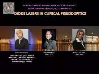
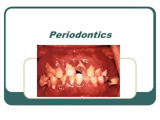
Lasers in Periodontics. Type of Lasers Laser Delivery Systems & Emission Modes Laser Interaction With Biologic Tissues Common dental Lasers & their Major Clinical Uses Advantages & Disadvantages of Laser Healing after laser therapy

E N D
Type of Lasers • Laser Delivery Systems & Emission Modes • Laser Interaction With Biologic Tissues • Common dental Lasers & their Major Clinical Uses • Advantages & Disadvantages of Laser • Healing after laser therapy • Lasers Safety in Dental Practice • References & Conclusion
L A S E R – is an acronym for light amplification by stimulated emission of radiation, is a form of electromagnetic energy in which photons are generated from a medium by stimulating the medium from external energy source. • The intraoral use of lasers has evolved over the last 4 decades as clinical experience along with scientific investigation, has increased the body of knowledge . • The dental lasers of today have benefited from decades of laser research and have their basis in certain theories from the field of quantum mechanics, initially formulated during the early 1900s by Danish physicist Bohr.
CLASSIFICATION OF LASERS: • There are many different types of lasers. The laser medium can be a solid, gas, liquid or semiconductor. Lasers are commonly designated by the type of lasing material employed. • Solid-State Lasershave lasing material distributed in a solid matrix (such as the ruby or neodymium: yttrium-aluminum garnet “Yag” lasers). The neodymium-Yag laser emits infrared light at 1,064 nanometers (nm). • Gas lasers(Helium and Helium- Neon, HeNe, are the most common gas lasers) have a primary output of visible red light. CO2 lasers emit energy in the far infrared, and are used for cutting hard materials.
Excimer lasers (the name is derived from the terms excited and dimers) use reactive gases, such as chlorine and fluorine, mixed with inert gases such as argon, krypton or xenon. When electrically stimulated, a pseudo molecule (dimer) is produced. When lased, the dimer produces light in the ultra violet range. • Dye lasersuse complex organic dyes, such as rhodamine 6G, in liquid solution or suspension as lasing media. They are tunable over a broad range of wavelengths. • Semiconductor lasers, sometimes called diode lasers, are not solid-state lasers. These electronic devices are generally very small and use low power. They may be built into larger arrays, such as the writing source in some laser printers or CD players.
CLASSIFICATIONS I. Based on application • Soft tissue laser eg: Argon, Co2, diode; Nd:YAG. • Hard tissue laser eg: Er : YAG • Resin curing laser eg: Argon • II. Mode of action • Contact mode (focused or defocused) • eg: Ho : YAG ; Nd: YAG • Non-contact mode (focused or defocused) • eg: CO2
III. Based on Level of energy emission: Soft lasers (low level energy): Athermal low energy lasers emitted at wave length, which are supposed to stimulate cellular activity. Example: He-Neon; Ga-Arsenide. Hard lasers (High level energy): Thermal lasers emitted at wavelength in the visible infra red and U.V range. Example: Er:YAG laser ; Nd: YAG laser. IV. Based on radiant energy generation: • Continuous wave or continuous form (CW) • Discreet or single pulses • Multiple timed pulses (Pulse modes)
EMISSION MODES Laser device can emit the light energy in one of 3 basic modes: • Continuous wave: the beam is emitted at one power level continuously as long as the device is activated by pressing the foot switch. • Gated pulse mode: there are periodic alterations of the laser energy being on and off, similar to a blinking light. This mode is achieved by the opening and closing of a mechanical shutter in front of the beam path of a continuous wave emission. • Free running pulsed mode. This mode is unique in that large peak energies of laser light are emitted for an extremely short time span, usually in micro seconds, followed by a relatively long time in which the laser is off.
Laser Delivery Systems Fiberoptic cable Articulated arm
Laser Energy & Tissue Temperature Effect of laser energy on tissue depends on: -Degree of temperature rise -Corresponding reaction of interstitial & intercellular water -Laser parameters like: Emission mode : Power (watts) : Time of exposure : Wave form : Pulse duration : Angulation of laser : Optical property of tissues
Thermal Effects of Laser on Tissue Tissue Temp (ºC) Observed Effect • 40-50 Hyperthermia • 60 Coagulation, Protein Denaturation • 70-90 Welding • 100 Vaporization • 200 Carbonization
LASER EFFECT ON TISSUES The light energy from a laser can have four different interactions with the target tissue. • Photochemical interaction. • Photo thermal interaction • Photo mechanical interaction • Photo electrical interaction
Co2 Laser • 10,600 nm • Both pulsed and continuous wave • Readily absorbed by water- very effective for soft tissue surgery • Advantage over Scalpel- strong hemostatic and bacterocidal effects
When applied to hard tissues produces severe thermal damage, such as • cracking, • melting, and • Carbonization (Sasaki et al 2002) • So its use has been limited to soft tissue surgery
Dental plaque removal and calculus carbonization when used in root surface (Tucker et al 1996) • Destruction of microbial colonies without inflicting undue damage to the root surfaces in defocused mode (Coffelt et al 1997) • Inhibition of periodontal tissue attachment by residual char layer in vivo (Gopin et al 1997) • Increased fibroblast attachment after root conditioning in pulsed defocus mode ( Crespi et al 2002) • No reported clinical studies on application of the CO2 laser in periodontal pockets
Nd:YAG laser • Free-running pulsed wave laser • wavelength of 1,064 nm • Low absorption in water • Energy scatters or penetrates to the biological tissue • Its photothermal effect is useful for soft tissue surgery • Produce relatively thick coagulation layer
Has been widely accepted in soft tissue surgery • 1990- FDA approved for soft tissue surgery • Easy to deliver by optical fiber contact tip • Easy insertion into the pockets • 1997- FDA approved for sulcular debridemnt, first approval of laser application in periodontal pocket by FDA • Not suitable for hard tissue ablation
AAP does not recommend the use of laser curettage (AAP 2006) • ALD approves the adjunctive use of lasers for curettage following conventional mechanical root debridement • Should be used as an adjunct rather than a primary instrument in pocket • Nd:YAG laser as a primary debriding instrument could not demonstrate the same or better clinical outcomes than with conventional SRP • Insufficient calculus elimination as well as occasional inevitable thermal damages of root surface
Laser was equally or more effective than SRP in reducing or inhibiting recolonization of specific bacterial species for pocket treatment (Horton & Lin 1992) • SRP plus laser treatment showed more reduction of periodontopathic bacterial levels compared to SRP alone, but root surface alterations were observed after laser treatment (Ben Hatit et al. 1996) • Significant reduction in clinical and microbiological parameters when CO2 laser, Nd:YAG laser and ultrasonic scaling were compared, but no significant difference between these groups (Miyazaki et al 2003)
Er:YAG laser • Solid-state laser that generates a light with a wavelength of 2,940 nm • Since the Er:YAG laser is well absorbed by all biological tissues that contain water molecules, this laser is indicated for both soft and hard tissue ablation
The free-running pulsed Er:YAG laser has already been used clinically for caries removal and cavity preparation • 1997- FDA approved for hard tissue treatment such as caries removal and cavity preparation
1999- for sulcular debridement • 2004- for osseous surgery • Recently introduced: - Erbium,Chromium-doped: Yttrium-Scandium-Gallium-Garnet (Er,Cr:YSGG) laser with 2,780 nm wavelength - Erbium-doped:Yttrium-Scandium-Gallium-Garnet (Er:YSGG) laser with 2,790 nm wavelength - are highly absorbed by OH ions than water molecules.
Soft tissue- photothermal evaporation • Hard tissue- microexplosion or water mediated explosive ablation • The excellent ablation effect of the Er:YAG laser of both soft and hard tissues has received a lot of attention in the field of periodontal therapy, and has been extensively researched.
Calculus removal ability comparable to ultrasonic scaling (Aoki et al 2000) • Complete calculus removal without thermal change of root surface (Folwaczny et al. 2000) • Clinically adequate debridement without carbonization or other side-effects of clinical relevance (Frentzen et al. 2002) • No major compositional or chemically deleterious changes on the root surface except for reduction of organic components (Sasaki et al. 2002) • Better conditions for fibroblast attachment (Schoop et al. 2002)
Diode lasers • A solid-state semiconductor laser that typically uses a combination of Gallium (Ga), Arsenide (Ar), and other elements such as Aluminum (Al) and Indium (In) to change electrical energy into light energy • wavelength range is about 800– 980 nm • The laser is emitted in continuous-wave and gated-pulsed modes • Usually operated in a contact method using a flexible fiber optic delivery system
Poorly absorbed in water, but highly absorbed in hemoglobin and other pigments • Excellent soft tissue surgical laser • FDA approved oral soft tissue surgery in 1995 and sulcular debridement in 1998 by means of a diode laser • Exhibits thermal effects • Tissue penetration of a diode laser is less than that of the Nd:YAG laser • Deeper coagulation and more charring on the surface
Diode laser did not produce any deleterious effect on the root surface (Kreisler et al 2001) • Diode laser was unsuitable for calculus removal and altered the root surface in an undesirable manner (Schwarz et al 2003) • Dry or saline-moistened root specimens resulted in no detectable alterations; however, blood-coated specimens showed severe damage depending on the irradiation conditions (Kreisler et al. 2002)
Alexandrite laser • A solid-state laser employing a gemstone called Alexandrite • In 1995 Rechmann & Henning first reported that the frequency-doubled Alexandrite laser • Remove dental calculus in a completely selective mode without ablating the underlying enamel or cementum • Further studies are required to demonstrate the safety and effectiveness of this laser in clinical usage
Waterlase Laser energy and water combination (Hydrokinetics™). : uses a cool-water spray to cut teeth without generating heat. : provides pinpoint accuracy for caries removal, root canals and gingival contouring. Advantages -Replaces Drill -No Heat -No Vibration -Sterilizes Tooth -Maximizes Bonding
Complete FDA clearance list: • Exposure of unerupted teeth • Fibroma removal • Frenectomy and frenotomy • Gingival troughing for crown impressions • Gingivectomy • Gingivoplasty • Gingival incision and excision • Hemostasis • Cutting, shaving, contouring and resection of oral osseous tissues (bone) • Excisional and incisional biopsies
Implant recovery • Incision and drainage of abscesses • Leukoplakia • Operculectomy • Oral papillectomies • Reduction of gingival hypertrophy • Soft tissue crown lengthening • Sulcular debridement (removal of diseased of inflamed soft tissue • Treatment of canker sores, herpetic and aphthous ulcers of the oral Mucosa • Vestibuloplasty
Method of Laser incision For gingivectomy using Nd: YAG and Co2 laser, the beam is directed such that nowhere in its course it hits the bone
EXCISIONAL BIOPSY WITH LASERS Nd-YAG LASER BEING USED FOR THE EXCISION OF THE HEMANGIOMA HEMANGIOMA IMMEDIATELY AFTER LASER SURGERY POST OPERATIVE HEALING AFTER 6-WEEKS
APTHOUS ULCER MANAGEMENT WITH LASER THERAPY APTHOUS ULCER PRETREATMENT APTHOUS ULCER AFTER BEING TREATED WITH LASER HEALED APTHOUS ULCER POST TREATMENT
Advantages of Laser Dentistry: • Cut and coagulate precisely, in one step • Eliminate collateral tissue damage • Minimize post-operative inflammation and discomfort • Clean, bloodless operating field • Radiant coagulation: seal blood vessels without cauterization via conductive heat or carbonization • Lasers seal lymphatic vessels thus leading to less post operative swellling • Greater comfort during and after surgery. • Homeostasis and reduced risk of blood borne pathogens • Rarely needs suture • High patient acceptance of treatment.
Controlled depth of laser penetration • Hemostasis provides excellent visibility-even down in the pocket • Reduce patient discomfort and the need for anesthetic • Laser incisions heal faster than electro-cautery But Scalpel has distinct advantage of • Low cost • Positive tactile perception • Heal more rapidly than laser
Disadvantages • Retinal burn if no protection. • Prolonged exposure to pulp causes irreversible pulp damage. • High cost. • Specially trained personnel required. • Chances of explosion. • Aerosol contamination- respiratory hazards.
Healing following laser therapy • Carruin & leumanen(1987)- suggested that laser created wounds heal faster and produce less scar tissue • Whereas the study by hylton(1986) and bvell indicated otherwise - laser irradiated wound heal slowly. - it also causes more initial tissue damage - initial tensile strength of laser wound was found to be less than scalpel wound - ultimate tensile strength remains comparable - use of low level laser is suggested to improve healing remains inconclusive till date
Nd: Yag and Co2 laser alters the root surface protein : mineral ratio and affects the ability of fibroblasts to attachment and the residual char formation delays wound healing • Er: YAG laser produces least surface damage and no melting, charring or carbonization as seen in Nd: Yag and Co2 laser .
Types of Laser Hazards • Eye : Acute exposure of the eye to lasers of certain wavelengths and power can cause corneal or retinal burns (or both). Chronic exposure to excessive levels may cause corneal or lenticular opacities (cataracts). • Skin : Acute exposure to high levels of optical radiation may cause skin burns; while carcinogenesis may occur for ultraviolet wavelengths (290-320 nm). • Chemical : Some lasers require hazardous or toxic substances to operate (i.e., chemical dye, Excimer lasers). • Electrical : Most lasers utilize high voltages that can be lethal. • Fire : The solvents used in dye lasers are flammable. High voltage pulse or flash lamps may cause ignition.
Laser safety glasses saline soaked pads Removal of laser plume With high volume laser filtration masks evacuator