Download
1 / 49
540 likes | 817 Views
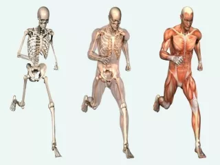
Development of the Musculoskeletal System. Objectives. At the end of this unit, the student will be able to: Discuss a timeline of normal development of the musculoskeletal system Discuss embryological development of bone, disc, muscle, tendon, joint, ligament and articular cartilage.

E N D
Objectives At the end of this unit, the student will be able to: Discuss a timeline of normal development of the musculoskeletal system Discuss embryological development of bone, disc, muscle, tendon, joint, ligament and articular cartilage.
Embryonic Development http://www.visembryo.com/baby/index.html
Notochord Development Solid cord of mesoderm cells Formed by the 18th day Midline, in cranio-caudal axis of the embryo First sign of skeletal formation Signals by secreting specialized proteins Signals overlying ectoderm to form the neural tube Eventually becomes the nucleus propulsus in the adult vertebrae
Germ Layer Development • Day 20: paraxial mesoderms form somites • Begins in cervical region, spread cranially and caudally • Cervical is most advanced part of body. • Three pairs formed per day until 44 pairs develop by week 5 • Some will later disappear
Somites • Somite pairs migrate • Cellular pairs • Ventromedial becomes Sclerotome (BONE) • Dorsolateral becomes Dermomyotome • Dermis, Dermatome • Myoblasts, Myotome
Axial structures - somites are arranged in pairs alongside the neural tube(light orange) and notochord (blue). Each somite is composed of a dermatome (light purple), myotome (light brown), and sclerotome (dark yellow). The ectoderm lies above and the endoderm below. • Somites, early on, become associated with a particular spinal nerve. • When many of the somite cells migrate to new locations in the embryo, they retain this neural link. • Basis for: • referred pain • patterns of cutaneousinnervation • nerve supply to muscles http://www.bionalogy.com/skeletal_system.htm
4 weeks • Major changes in body of embryo • Two pharyngeal arches are visible and two develop • Mandibular arch (first) gives rise to the lower jaw, extension of this gives rise to the upper jaw • Second arch is the hyoid arch • Upper limb buds are recognizable • Pouches for arms • UE forms before LE
5th week Enlargement of head caused by brain development Articular Cartilage Development Carnegie Development do not correspond to time, but rather a stage. Do Not Need to Know
6th week Upper limbs begin to show differentiation into the elbows and large handplates develop Primorida of the digits develop within the handplates Beginning of the development of the lower limbs Trunk and neck begin to straighten One of the most important times in Musculoskeletal development
6th Week= Vertebrae Development 6th week: chondrification centers appear in each mesenchymal vertebra Centers in each centrum fuse to form a cartilaginous centrum Centers in neural arches fuse with each other and cartilaginous centrum and spinous processes, transverse processes develop
6th week Neural tube(light orange) Notochord (blue)=NuclueosPulpuses Dermaome (light purple)=Vertebra Myotome (light brown) Sclerotome (dark yellow). http://www.bionalogy.com/skeletal_system.htm Intervertebral Disks develop from densely arranged cells which migrate cranially Remainder of densely packed cells fuse with loose cells to from a mesenchymal centrum. (primordium of vertebral body) Each centrum develops from two adjacent sclerotomes
7th week • Limbs undergo change=enlogation • Notches appear between the digits • By end of the week, ossification of the bones in the upper limb has begun • Clavicle one of the first
8th week Digits of the hand are separated but still webbed. Notches visible between digital rays of the feet All digits have lengthened and are completely separated Ossification begins in the femur
9-12 weeks Head is half the length of the body 9 weeks: legs are short, get longer but not to the full extent as at birth, arms are almost their normal length Primary ossification centers appear in skeleton by end of 12 weeks
Specific Tissue Formation • Cartilage • Bone • Vertebrae • Sternum • Ribs • Muscle Tendon Joints Ligaments Capsule
Development of Axial Skeleton • During fourth week • Mesenchymal cells from the sclerotomes are around the notochord, neural tube and in body wall • Sclerotome: loosely arranged cells cranially, densely packed cells caudally
4th week • Notochord degenerates and disappears in areas that are surrounded by the developing vertebral bodies • Above the vertebral bodies, notochord develops into the nucleus pulposus Snell, R
Development of Bone and Cartilage • Mesoderm gives rise to mesenchyme: meshwork of loosely organized embryonic connective tissue • Bones appear as condensations of mesenchymal cells to form a bone model
Mesenchymal Bone Formation • Intramembranous ossification • Occurs in mesenchyme that has formed a sheath, becomes highly vascularized • Develops into osteoblasts which deposit osteoid Snell R. Clinical Embryology for Medical Students, 2nd ed. 1972, Little Brown and Co. Boston, MA
Process of Bone Formation Osteoid is cell matrix, deposited by the mesenchyme cells. Calcium phosphate attaches to the osteoid tissue and traps the osteoblasts within. These cells then become osteocytes. No pattern develops at first, later organize into lamellae which develop around blood vessels. Osteons (Haversian system)
Osteoblasts at periphery of developing bone, lay down the lamella, forms plates of compact bone with spongy bone in between • Between spongy environment is region of osteoclastic activity (bone resorption) • Mesenchyme also develop into bone marrow in this region Snell, R
Cartilage Model of Bone Formation • Endochondral Ossification • Primary Center of Ossification • Give the initial length to bone • Secondary Centers of Ossification • Growth plates Junqueira LC, Carneiro J.
Primary Ossification Center • Appears in diaphysis • Chondrocytes increase in size by mitosis, align in rows, enlarge to increase length and diameter • Chondrocytes then die, matrix calcifies and forms a honeycomb appearance • Thin layer of bone deposited under perichondrium which becomes periosteum • Process moves toward the epiphyses
At Birth: diaphyses are largely ossified • Epiphyses are still cartilagenous • Secondary ossification centers occur within a few years after birth Junqueira and Carneiro
Summary of Bone Growth http://en.wikipedia.org/wiki/Image:Illu_bone_growth.jpg
Vertebral Ossification • Ossification of vertebrae begins during embryonic period and ends by age 25 • Ossification within neural arches by eighth week • Birth: each vertebrae has three bony parts connected by cartilage • Fuse within first 3-5 years • Sometimes the vertebrae will fuse together. Snell, R
6th week Snell, R
Arches unite first in lumbar region and progress cranially • Vertebral arch articulates with the centrum at cartilaginous neurocentral joints. These joints permit the vertebral arches growth as the spinal cord enlarges • Five secondary ossification centers appear after puberty – ossify by age 25: • Tip of spinous process • Tip of each transverse process • Superior and inferior vertebral end plates
Exceptions to typical vertebral ossification • C1 vertebrae • C2 vertebrae • C7 vertebrae • All lumbar, sacral and coccyx vertebrae • Typical: • 7 cervical vertebrae • 12 thoracic • 5 lumbar • 5 sacral • Sometimes L5 fuses to the sacrum (sacralization) • Sometimes S1 doesn’t fuse (lumbarization)
Development of the Disc Mesenchyme cells form the annulus fibrosus Notochord forms the nucleus pulposus Sclerotome cells migrate medially to surround the notochord Notochord becomes encased in a continuous cylinder of sclerotomicmesenchyme Caudal half of each sclerotome fused with cephalic half of the next scelrotome to form the mesenchymal vertebral body 4th week of gestation:
Ghosh P: The Biology of the Intervertebral Disc, Vol. I, CRC Press, 1988 5-6 weeks gestation: mesnechymal tissue surrounding notochord begins to segment Mesenchymal condensations forms the annulus fibrosus (light band) Non condensed mesenchyme forms the vertebral bodies (dark band)
Notochord enlarges between vertebral bodies, forms nucleus propulsus Notochord disappears in areas where vertebrae are developing
Development of Ribs Ribs develop from mesenchymal costal processes of thoracic vertebrae Cartilaginous during embryonic period Ossify during fetal period Seven pairs true ribs Five pairs of false ribs Two pair of floating ribs
Development of Sternum Pair of sternal bars develop ventrolaterally in body wall Condrification occurs in bars as they move medially Fuse craniocaudally to from cartilaginous models of manubrium, sternum and xiphoid process Centers of ossification appear craniocaudally in sternum before birth except for xiphoid process which occurs during childhood
Development of Limbs Fifth Week: condensations of mesenchyme appear as limb buds Mesenchymal bone models in limbs chondrify during the sixth week and form hyaline cartilage bone models Clavicle develops first then pectoral girdle and upper limb bones then pelvic girdle and lower limb bones
Ossification begins by 8th week and occurs in primary ossification centers 12th week, all long bones have primary ossification centers Clavicles begin to ossify before any other bone in the body, then the femurs Secondary ossification centers occurs in utero, first in the knee, most appear after birth
Early 7th week: limbs extend ventrally. Flexor aspect of limbs are ventral and extensors are dorsal. • UE rotate Externally • LE rotate Internally • Limbs then rotate in opposite directions and to different degrees • Upper limbs rotate laterally through 90 dg • Elbows face posterior • Flexors of elbow are anterior • Lower limbs rotate medially through almost 90 dg • Knees face anterior • Extensors of knee are anterior
Embryological Development of Joints Develop during the sixth week with the appearance of the interzonalmesenchyme. By the end of week eight, they resemble adult joints. Layer of synovium separates from the joint spaces
Fibrous Joint Development Dense fibrous tissue develops from the interzonalmesenchyme creating the fibrous joints like the cranial sutures.
Cartilaginous Joint Development • Interzonal mesenchyme develops into either hyaline cartilage (chostochondral joints) or fibrocartilage (pubic symphysis, clavicle) • Starts week 6 continues into week 8
Synovial Joint Development • Interzonal mesenchyme differentiates into (week 7-8): • Peripherally, it forms the capsular and other ligaments • Centrally it disappears and resulting space becomes the joint cavity or synovial cavity • When it lines the joint capsule and articular surfaces, it becomes the synovial membrane
Development of Skeletal Muscle Derived from the myotome regions of somites First indication of myogenesis is the elongation of the nuclei and cell bodies of the mesenchymal cells which differentiate into myoblasts. When start to see striation then it is differeante.
The primordial muscle cells fuse to form elongated, multinuclear, cylindrical tubes or myotubes. Muscle growth in the fetus then occurs with ongoing fusion of myoblasts and myotubes.
At the time of the fusion myofilaments appear in the cytoplasm of the myotubes Most skeletal muscle is developed before birth All developed by age 1, muscles increase in length and width as the skeleton grows.
Embryological Development of Tendon Embryologically, tendon develops from the mesoderm tissue Develops at a similar time as ligaments and muscle Initially consists of tenoblasts and type I collagen
Upper limb tendons develop before lower limb tendons Extensor muscles of lower limb don’t fully develop until after birth Interaction occurs between the tendon cells and muscle cells to coordinate correct insertion of each tendon to its appropriate muscle (Jozsa, Kannus, 1997)
Development of Ligament:EX: Anterior Cruciate Ligament Weeks 9-10 Fibroblasts align between femoral and tibial attachment sites Collagen fibrils are deposited between cells, parallel to the longitudinal axis of the ligament As cells deposit more and more collagen, bundles of matrix push cells apart forming a mature ligament with a low density of cells and a higher collagen content
References Moore KL, Persaud TVN. The Developing Human. Clinically Oriented Embryology, 8th ed. Saunders Elsevier, Philadelphia PA, 2008. Cech DJ, Martin S. Functional Movement Development Across the Life Span, 2nd ed. New York, W.B. Saunders. 2002 Whitaker R. Embryology Lectures downloaded from www.instantanatomy.net , Podcast accessed on April 30, 2011.