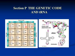
Section P Genetic code and tRNA
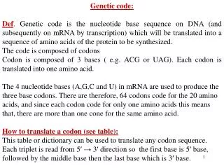
Section P Genetic code and tRNA. P1 The genetic code P2 tRNA structure and function. P1 The genetic code. Nature Deciphering Feature Effect of Mutation Universality ORFs Overlapping Genes. Nature. Genetic code is a triplet code (to be grouped in 3nt);

Section P Genetic code and tRNA
E N D
Presentation Transcript
Section P Genetic code and tRNA P1 The genetic code P2 tRNA structure and function Yang Xu, College of Life Sciences
P1 The genetic code • Nature • Deciphering • Feature • Effect of Mutation • Universality • ORFs • Overlapping Genes Yang Xu, College of Life Sciences
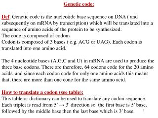
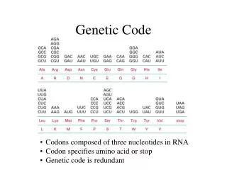
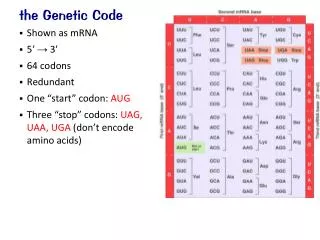
Nature • Genetic code is a triplet code (to be grouped in 3nt); • The triplet codons are adjacent (non-overlapping); • They are not separated by punctuation (comma-less). • Because many of the 64 codons specify the same amino acid, the genetic code is degenerate; • So that the genetic codons have redundancy. As more gene and protein sequence information has been obtained, it has become clear that the genetic code is very nearly, but not quite, universal. This supports the hypothesis that all life has evolved from a single common origin. Yang Xu, College of Life Sciences
Deciphering • In the 1960s, Marshall Nirenberg developed a “cell-free protein synthesizing system” from E. coli (细胞外蛋白合成体系). • To determine which amino acids were being polymerized into polypeptides, it was necessary nucleotide phosphorylase was used to make synthetic mRNAs that were composed of only one nucleotide, that is poly (U), poly (C), poly (A) and poly (G). • If protein synthesis took place after adding one of these mRNAs, then in one of the 20 reaction tubes, the radioactivity could show the formation of polypeptide. • In this way, it was found that: • Poly (U) caused the synthesis of poly-phonylalanine, • Poly (C) coded for poly-proline • Poly (A) for poly-lysine • Poly (G) did not work because it formed a complex secondary structure. Yang Xu, College of Life Sciences
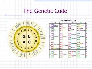
Deciphering Yang Xu, College of Life Sciences
Deciphering • The precise sequence of the triplet codon can only be worked out if additional information is available. • Towards the end of the 1960s, it was found that synthetic tri-nucleotides could attach to the ribosome and bind their corresponding aminoacyl-tRNAs. • Upon filtering through a membrane, only the complex of ribosome, synthetic triplet and aminoacyl-tRNA was retained on the membrance. Yang Xu, College of Life Sciences
Feature • The genetic code is degenerate (or it shows redundancy). This is because 18 out of 20 amino acids have more than one codon to specify them, called synonymous codons . • Only methionine and tryptophan have single codons. • The synonymous codons are not positioned randomly, but are grouped in the table. Generally they differ only in their third position. • In all cases, if the third position is a pyrimidine, then the codons specify the same amino acid (are synonymous). • In most cases, if the third position is a purine the codons are also synonymous. Yang Xu, College of Life Sciences
Modifications of the genetic code Codon Usual Alternative Organelle/organism AGA Some Animal AGG Mitochondria AUA Ile Met Mitochondria CGG Arg Trp Plant Mitochondria CUN Leu Thr Yeast Mitochondria AUU Ile GUG Val Start Some Prokaryotes UUG Leu UAA UAG UGA Stop Trp Mit. Mycoplasma Arg Stop, Ser Stop Glu Some Protozoans P254, P1.2 Universality • For a long time after the genetic code was deciphered, it was thought to be universal, that is the same in all organisms. • However, since 1980, it has been discovered that mitochondria, which have their own small genomes, use a genetic code that differs slightly from the standard, or ‘universal’ code. Indeed, it is now known that some other unicellular organisms also have a variant genetic code. Yang Xu, College of Life Sciences
Overlapping Genes • Generally overlapping genes occur where the genome size is small and there is a need for greater information storage density. • In viruses, for example, the phage ΦΧ174 makes 11 proteins of combined molecular mass 262 kDa from a 5386 bp genome. Without overlapping genes, this genome could encode at most 200 kDa of protein. Three proteins are encoded within the coding regions for longer proteins. • In prokaryotes, the ribosomes simply have to find the second start codon to be able to translate the overlapping gene and they may achieve this without detaching from the template. • Eukaryotes have a different way of initiating protein synthesis and tend to make use of alternative RNA processing to generate variant proteins from one gene. Yang Xu, College of Life Sciences
Robert Holley(46y) P2 tRNA Structure and Function • tRNA Primary Structure • tRNA Secondary Structure • tRNA Tertiary Structure • tRNA Function • Aminoacylation of tRNA • Aminoacyl-tRNA Synthetases • Proofreading Yang Xu, College of Life Sciences
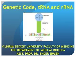
tRNA Primary Structure • tRNAs are the adaptor molecules that deliver ammo acids to the ribosome and decode the information in mRNA. Their primary structure (i.e. the linear sequence of nucleotides) is 60-95 nt long, but most commonly 76 nt. • They have many modified bases sometimes accounting for 20% of the total bases in any one tRNA molecule. Indeed, over 50 different types of modified base have been observed in the several hundred tRNA molecules characterized to date, and all of them are created post-transcriptionally. • Seven of the most common types are shown in Fig. 1 as nucleosides. Yang Xu, College of Life Sciences
tRNA Secondary Structure • All tRNAs have a common secondary structure (i.e. base pairing of different regions to form stems and loops), the cloverleaf structure. Yang Xu, College of Life Sciences
34 ---aa accept arm loading aa at 3’ end ---TΨC loop: contact with 5s rRNA ---DHU loop: contact with AARS ---anti-codon loop: 34th is wobble base ---extra loop: (Variable arm)classification marker ? Yang Xu, College of Life Sciences
tRNA Tertiary Structure • There are hydrogen bonds (tertiary hydrogen bonds) that help form the 3-D structure of tRNA molecules. Yang Xu, College of Life Sciences
tRNA Function • tRNAs are joined to amino adds to become aminoacyl-tRNAs (charged tRNAs) in a reaction called aminoacylation. • It is these charged tRNAs that are the adaptor molecules in protein synthesis. • Special enzymes called aminoacyl-tRNA synthetases carry out the joining reaction which is extremely specific (i.e. a specific amino acid is joined to a specific tRNA). • These pairs of specific amino acids and tRNAs, or tRNAs and aminoacyl-tRNA synthetases are called cognate pairs, and the nomenclature used is shown in Table 1 (page 258). Yang Xu, College of Life Sciences
Aminoacylation of tRNA • The general aminoacylation reaction is shown in Fig. It is a two-step reaction driven by ATP: • In the first step, AMP is linked to the carboxyl group of the amino acid giving a high-energy intermediate called an aminoacyl adenylate. • The hydrolysis of the pyro-phosphate released (to two molecules of inorganic phosphate) drives the reaction forward. • In the second step, the aminoacyl adenylate reacts with the appropriate uncharged tRNA to give the aminoacyl-tRNA and AMP. Yang Xu, College of Life Sciences
Aminoacyl-tRNA Step 1 Setp 2 Yang Xu, College of Life Sciences
Aminoacyl-tRNA Synthetases • Despite the fact that they all carry out the same reaction of joining an amino acid to a tRNA, the various synthetase enzymes can be quite different. • They fall into one of four classes of subunit structure, being either a, a2, a4, a2b2. • The polypeptide chains range from 334 to over 1000 amino acids in length, and these enzymes contact the tRNA on the underside (in the angle) of the L-shape. • They have a separate amino acid-binding site. The synthetases have to be able to distinguish between about 40 similarly shaped, but different, tRNA molecules in cells, and they use particular parts of the tRNA molecules, called identity elements, to be able to do this (Fig. 4). Yang Xu, College of Life Sciences
Identity Elements (Paracodon) AARS tRNA binding site aa binding site Paracodon of tRNA loading Amino Acid(R) Yang Xu, College of Life Sciences
tRNAPhe tRNAAla Identity Elements in various tRNA Molecules tRNAfMet tRNASer Yang Xu, College of Life Sciences
aa arm: A,D,G,H,N,S,T,V,W TΨC loop 73th site A.C.D.E.F.G.H.I.K.L.M.N.P.Q.R.S.V.W.Y. Extra loop A, F, L, R Anti-codon loop C,D,E,F,G,H,I,K,M,N,PQ,R,T,V,W,Y Position of Identity Elements Yang Xu, College of Life Sciences
That’s all for Section P Yang Xu, College of Life Sciences