Download
1 / 21
240 likes | 957 Views
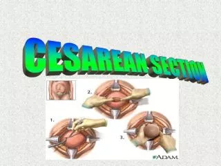
Cesarean Section Scar Defects (CSDs). Dr Muhammad M El Hennawy Ob/gyn Consultant Rass el barr –dumyat - egypt www.mmhennawy.co.nr. Cesarean Scar Defects (CSDs). It is a deficient uterine scars or scar dehiscence following a cesarean section, involve myometrial discontinuity

E N D
Cesarean Section Scar Defects (CSDs) Dr Muhammad M El HennawyOb/gyn ConsultantRass el barr –dumyat - egyptwww.mmhennawy.co.nr
Cesarean Scar Defects (CSDs) It is a deficient uterine scars or scar dehiscence following a cesarean section, involve myometrial discontinuity at the site of a previous cesarean section Scar Symptomatic cesarean scar defect is one of the commonly reported long-term complications of cesarean section
Incidence • In the literature varies widely, • Ranging from 0.3% to 19.4% • (prolonged postmenstrual spotting was the most common (63.8%) followed by dysmenorrhea (53.1%), chronic pelvic pain (39.6) and dyspareunia (18.3%,)).
Shapes • a pouch on the anterior uterine segment at the site of the cesarean scar different shapes such • Thin linear defect, • Focal saccularoutpouching, • Unilateral or bilateral diverticula (dog-ear like) and • Fistula and • Different locations such as the uterine body, lower uterine segment, uterine isthmus and the upper endocervical canal
A Histopathological Study Of Hysterectomy Specimens With Cesarean SectionScars Three possible mechanisms underlying the pathogenesis of this condition: Firstly, The presence of a congested endometrial fold (61%) and small polyps in the scar recess (16%)) are potential causes of menorrhagia and abnormal uterine bleeding; Secondly, Lymphocytic infiltration (65%)) and distortion of the lower uterine segment (75%) could contribute to chronic pelvic pain and dyspareunia; Thirdly, Iatrogenic adenomyosis confined to the scar (28%) could account for dysmenorrhea.
Measurements • D=depth • W=width • T = residual myometrium • The myometrial defects were measured and evaluated with regard to the following parameters: • Scar width (the length of the widest gap along the cervicoisthmic canal), the mean width was 4.575mm, • Scar depth (the vertical distance between the base and apex of the defect) mean depth 4.295mm, and • Thickness of the residual myometriumthe mean myometrial thickness overlying the scar was 1.314mm, and the mean myometrial thickness adjacent to scar was 4.533 mm • .
Risk Factors for Incomplete Healing of the Uterine Incision After Caesarean Section • Cesarean section in advanced labor increases the risk of incomplete healing of the uterine incision. • Multiple cesarean section as repeated trauma to a wound can disrupt the normal healing process • PROM • Surgical technique used to close the uterine incision eg1- versus 2-layer closure,includedecidua in sutures and locking sutures • increasing gestational week at delivery, • Pre-eclampsia • Blood loss, • Cervical dilatation • The station of the presenting fetal part at the time of delivery • Duration of labor • Oxytocin augmentation • Retroflexed uterus
Surgical Technique Used To Close The Uterine Incision • 1 - The number of layers, Closure of the transverse uterine incision using a single running locking suture penetrating the full thickness of the myometrium and endometrium has been associated with a two- to four-fold risk of uterine rupture compared to double-layer closure. • 2 – The inclusion or exclusion of the endometrium in the suture, a single-locked suture of the uterus is secondary to the fact that this technique is usually performed with inclusion of the decidua(endometrium) in the scar tissue. • 3 - The locking (or not) of the suture. the single-layer closure was performed in a locking fashion , it is possible that the locked suture, by being more hemostatic, can cause a strangulation of the scar tissue and lead to weaker healing
Long-Term Complications Of Cesarean Section = During pregnancy - Ectopic pregnancy (cesarean section scar pregnancy)CSP - Early placenta accreta (pathologically adherent placenta)EPA - Placenta previa = During pregnancy ,abortion and labour - Ceesarean scar dehiscence following incomplete abortion secondary to uterotonic medication - Scar dehesience - Scar rupture = Non pregnant - Abnormal uterine bleeding ( postmenstrual bleeding-metrorrhagia) due to the presence of a congested endometrial fold and small polyps in the scar recess or The proposed mechanism of abnormal uterine bleeding is a pouch or “isthmocele” in the lower uterine segment that causes delayed menstrual bleeding or Uterine scar diverticuli may cause intermenstrual bleeding -Chronic pelvic pain , dull sensation following menstruation , dyspareunia, due to lymphocytic infiltration and distortion of the lower uterine segment - Dysmenorrhea due to iatrogenic single large focal adenomyosis confined to the uterine scar -Unexplained infertility = as well as a potentially higher risk of complications and difficulties during gynecologic procedures such as uterine evacuation, hysterectomy, endometrial ablation, and insertion of an intrauterine device.
The Systematic Correction Of A Dehiscence Before A Pregnancy It is probably not indicated. Nevertheless, since the risk of rupture is at its greatest during labor the rate of uterine rupture or dehiscence is on average 6.6% (range 1% to 46%) Thus performing an elective Cesarean section before the onset of labor in these patients. Another guidelines for the management of women contemplating a VBAC, with a very low rate of uterine rupture using a cut-off value of 3.5 mm measured transabdominal or tranvaginal
Transvaginal Sonography • It is a very simple, noninvasive, low-cost examination that should be considered as the first choice for screening, because it highly correlates (100%) with hysteroscopy in the diagnosis of this defect and may help rule out other causes.
Saline Contrast Sonohysterography • Niches can be identified by SCSH (saline contrast sonohysterography )following a Cesarean section in about 60% of patients. • The prevalence of scar dehiscence (6%) is much higher than the reported risk of uterine rupture (0.4%). In non-pregnant women CS scars are better evaluated at SCSH than at unenhanced ultrasound examination, because the demarcations of scar defects are more clearly delineated at SCSH than before. Moredefects were detected and more defects were classified as large at SCSH
Laparoscopic excision (Laparoscopic repair of the dehiscence, including excision of the fibrotic tissue and laparoscopic closure of the anterior uterine wall) • Resectoscopic treatment (Hysteroscopic resection of fibrotic tissue that overhangs underneath the triangular pouch showing the muscular tissue below, facilitating blood drainage through the cervix and fulguration of endometrial glands and/or dilated blood vessels), • Combined laparoscopic and vaginal scar excision and uterine repair • Vaginal revision, and • Endometrial ablation.
Laparoscopic Repair of Cesarean Section Scar Defects in Nonpregnant Women • A) Laparoscopic view of the cesarean scar with a probe inserted into the endocervix. The residual myometrium covering the scar is very thin (arrow). • (B) Laparoscopic view of the cesarean section scar defect cavity (arrows). • (C) Laparoscopic view of the first layer of suture (arrows). • (D) Laparoscopic view of the second layer of suture (arrows).
Resectoscopic treatment • Hysteroscopy identified the pouch as a cleft on the anterior wall of the cervix just at the end of the cervical channel (after the internal os), in correspondence with the defect shown on transvaginalsonography • Surgical treatment under hysteroscopic vision for resecting the fibrotic tissue that sometimes overhangs below the scar, thereby improving menstrual drainage and avoiding blood accumulation
Cesarean Delivery Scar Ectopic Pregnancy • Cesarean scar pregnancy should be diagnosed and treated as soon as possible to prevent severe complications and spare fertility. • Cesarean scar pregnancy is a rare diagnosis but should always be considered in a patient with a low-lying gestational sac and an appropriate surgical history. • Key sonographic criteria may help distinguish cesarean scar pregnancy from the 2 most likely differential diagnoses--spontaneous abortion in progress and cervical ectopic pregnancy • the following sonographic findings should raise the suspicion level for a Cesarean Scar Pregnancy: -No fetal parts in the uterine cavity or cervix -A thin myometrial layer between the bladder and gestational sac -A triangular-shaped gestational sac -A gestational sac that is close to the bladder and uterine wall -Presentation of arteriovenous malformation in the area • systemic methotrexate therapy, local injection of methotrexate or other embryocides, gestational sac aspiration, dilatation and curettage, surgical laparotomy/hysterotomy, hysteroscopy, laparoscopy, and uterine artery embolization. • However, many feel that expectant management and blind dilatation and curettage should be avoided as significant complications have been reported. • Ultimately, the approach depends on various factors such as gestational age at presentation, hemodynamic stability, local endoscopic expertise, future fertility plans, and feasibility of serial follow-up serology and imaging.
Laparoscopy Surgical Treatment of Caesarean Scar Ectopic Pregnancy (CSEP) • Step 1: Cystoscopywith bilateral ureteral catheterization and Laparoscopy shows a mass arising from the serosa was detected at the level of the scar of the cesarean section. • Step 2: Hemorrhage Control by temorary closure of the internal iliac arteries by sutures • Step 3: Monobloc resection and correction of the scar defect The uterine serosa was incised to isolate the bladder , Elliptical excision of the mass containing geststional sac and the affected uterine wall ,removed en bloc , the uterine defect was repaired using 2-0 p (Vicryl) in two planes with continuous suture and 3-0 polydioxanone (PDS II) suture for closing the serosa
Vaginal Temporary occlusion of uterine artery • Dilatation evacuation curretage • Foley balloon inflation in cervix • Remove temporary occlussion of UA
DD Of CSDs By HSG • In the interpretation of a hysterosalpingogram, awareness of the appearance of the cesarean scar defect is important to avoid misdiagnosis of the scar as underlying pathology or normal variants • 1-Prominent cervical glands, small tubular structures arising from the cervical wall, which are typically multiple, bilateral and symmetric unlike the cesarean scar • 2-Nabothian cyst that is commonly seen in the stroma of the cervix • 3-Post myomectomydiverticulum; small outpouching at the resection site other than the cesarean section scar depending on the surgical site. The history may also be helpful. • 4-Post curettage diverticulum. The patient’s clinical history is important. A previous dilatation and curettage (D&C) history is helpful • 5-Congenital cervical diverticula. If the patient has no surgical history, it is helpful in the differential diagnosis • 6-Focal adenomyosis: Ingrowing of the endometrial tissue into the myometrium with adjacent smooth muscle hyperplasia. It is seen in HSG as fine channels extended perpendicular to the uterine cavity ending in small diverticulum-like structures; focal adenomyosis are multiple and smaller than the cesarean section scar and accompany uterine enlargement, while cesarean section scars are usually larger and single • 7-Tuberculosis: No cesarian section history and obstruction of the fallopian tubes are helpful