Download
1 / 108
1.1k likes | 1.79k Views
Myocardial Infarction and its Complications. REZA KIANI ,MD, Invasive Cardiologist & Interventionist Rajaei Heart Center. Introduction. Coronary artery disease is the leading cause of mortality in modern societies and is a worldwide epidemic.

E N D
Myocardial Infarction and its Complications REZA KIANI ,MD, Invasive Cardiologist & Interventionist Rajaei Heart Center
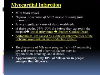
Introduction • Coronary artery disease is the leading cause of mortality in modern societies and is a worldwide epidemic. • In 2001, it was estimated that worldwide, ischemic heart disease was responsible for 11.8 percent of all deaths (5.7 million) in low-income countries and 17.3 percent (1.36 million) of all deaths in high-income countries. • Acute Myocardial Infarction (MI) is the most common cause of fatality in the family of coronary artery disease. • Despite revolutionary improvements in the treatment of this catastrophic disease, AMI continues to be a serious public health problem • it has been estimated that the number of years of life lost because of an AMI is 14.2 years. • the cost to American society (both direct and indirect) is $142.5 billion per year.
Mortality rate • Observational data bases suggest that 30-day mortality rate in MI patients in the community is 15 to 20 percent . • The mortality rate of patients with STEMI who receive an appropriate pharmacological or invasive therapy is in the range of 5.5 to 7.5 percent. • So, appropriate therapy causes 9-12% improvement in survival in 30 days. Impressive!!
Definition of MI • The pathological diagnosis of myocardial infarction (MI) :evidence of myocyte cell death as a consequence of prolonged ischemia. Characteristic findings include coagulation necrosis and contraction band necrosis, often with patchy areas of myocytolysis at the periphery of the infarct. • the majority of myocyte loss in the infarct zone occurs via coagulation necrosis and proceeds to inflammation, phagocytosis of necrotic myocytes, and repair eventuating in scar formation.
The clinical diagnosis of MI requires an integrated assessment of the history& physical exam with some combination of indirect evidence of myocardial necrosis using biochemical, electrocardiographic, and imaging modalities .
Pathology • During the natural evolution of atherosclerotic plaque, especially lipid laden plaque, an abrupt transition can occur, characterized by plaque disruption . • Plaque disruption exposes substances that promote platelet activation and aggregation, thrombin generation, and ultimately thrombus formation. • The resultant thrombus interrupts blood flow and leads to an imbalance between oxygen supply and demand and, if this imbalance is severe and persistent, to myocardial necrosis
Why an atherosclerotic plaque ruptures? • Activated macrophages and mast cells abundant at the site of atheromatous plaque are an important cause of rupture. These cells overexpressmetalloproteinase enzymes such as collagenase, gelatinase, and stromelysinthat degrade components of the protective extracellular matrix such as collagen, elastin and glycoproteins. • In addition, stresses induced by intraluminal pressure, tachycardia (cyclic stretching and compression), and disruption of nutrient vessels combine to produce plaque disruption . • Plaques always disrupt at the margin of the fibrous cap near an adjacent, less involved segment of the coronary artery wall (shoulder region of plaque)
After disruption • Platelet aggregation to the surface of ulcerated plaque is the first step of thrombus formation • After that, the platelets release ADP, thrombin, serotonin, epinephrinwhich causes fibrin formation, entrapment of red blood cells and propagation of the early thrombus.
The composition of the thrombus may vary at different levels: A white thrombus is composed of platelets, fibrin, or both, and a red thrombus is composed of erythrocytes, fibrin, platelets, and leukocytes. • Early thrombi are usually small and non-occlusive and are composed predominantly of platelets. • Occlusive thrombi and propagation of early thrombus are predominantly red thrombus.
Gross pathology • On gross inspection, myocardial infarction can be divided into two major types: transmural infarcts, in which myocardial necrosis involves the full thickness (or nearly full thickness) of the ventricular wall, and subendocardial (nontransmural) infarcts, in which the necrosis involves the subendocardium and inner part of intramural myocardium, without extending all the way through the ventricular wall .
An occlusive coronary thrombosis appears to be far more common when the infarction is transmural . • Nontransmural infarctions, however, frequently occur in the presence of severely narrowed but still patent coronary arteries.
Gross pathology cont. • THE FIRST HOURS. • Gross alterations of the myocardium are difficult to identify until at least 6 to 12 hours have elapsed following the onset of necrosis • THE FIRST DAYS. • Eighteen to 36 hours after the onset of the infarct, the myocardium is reddish purple (because of trapped erythrocytes), with a serofibrinousexudateevident on the epicardium in transmural infarcts. These changes persist for approximately 48 hours; the infarct then turns gray, and fine yellow lines, secondary to neutrophilic infiltration, appear at its periphery. This zone gradually widens and during the next few days extends throughout the infarct. • THE FIRST WEEKS. • Eight to 10 days after infarction, the thickness of the cardiac wall in the area of the infarct is reduced as necrotic muscle is removed by mononuclear cells. The cut surface of an infarct of this age is yellow, surrounded by a reddish purple band of granulation tissue that extends through the necrotic tissue by 3 to 4 weeks. Commencing at this time and extending over the next 2 to 3 months, the infarcted area gradually acquires a gelatinous, ground-glass, gray appearance, eventually converting into a shrunken, thin, firm scar, which whitens and firms progressively with time
Patterns of myocardial necrosis • COAGULATION NECROSIS. • Coagulation necrosis is usually present in the central region of infarcts, which results in the arrest of muscle cells in the relaxed state and the passive stretching of ischemic muscle cells. no calcification is evident. • NECROSIS WITH CONTRACTION BANDS. • This form of myocardial necrosis, also termed contraction band necrosis or coagulativemyocytolysis, results primarily from severe ischemia followed by reflow.It is characterized by hypercontracted myofibrils with contraction bands and mitochondrial damage, frequently with calcification. Necrosis with contraction bands is caused by increased Ca2+ influx into dying cells, resulting in the arrest of cells in the contracted state. It is seen in the periphery of large infarcts and is present to a greater extent in nontransmuralthan in transmural infarcts. • MYOCYTOLYSIS. • Ischemia without necrosis generally causes no acute changes that are visible by light microscopy. However, severe prolonged ischemia can cause myocyte vacuolization, often termed myocytolysis. Prolonged severe ischemia causes hydropic, vascular, and fatty degeneration.
polymorphonuclearleukocytic infiltrate in an area of acute myocardial infarction of 3 to 4 days' duration Nearly complete removal of necrotic myocytes by phagocytosis (â≈7 to 10 days).
Granulation tissue with a rich vascular network and early collagen deposition, approximately 3 weeks after infarction replacement of the necrotic fibers by dense collagenous scar
Effect of reperfusion ,either spontaneous or as part of therapy
Pathophysiology • Systolic function: Upon interruption of antegrade flow in an epicardial coronary artery, the zone of myocardium supplied by that vessel immediately loses its ability to shorten and perform contractile work. • 3 abnormal contraction patterns develop in sequence: (1) hypokinesis, reduction in the extent of shortening; (2) akinesis, cessation of shortening; and (3) dyskinesis, paradoxical expansion, and systolic bulging. • If a sufficient quantity of myocardium undergoes ischemic injury, left ventricular pump function becomes depressed; cardiac output, stroke volume, blood pressure. • Reducedcardiac output causes: hypotension & shock, hypoperfusion of visceral organs ( kidney, GI tract, Liver) and if severe enough perfusion of heart and CNS also will decrease.
Diastolic function: • relaxation of heart muscle is an active and energy-dependent process. So, in ischemia, relaxation of the heart is compromised just like the contraction . • impaired diastolic function causes backward heart failure : increased LV diastolic pressure increases pulmonary artery venous pressure and capillary pressure( pulmonary wedge pressure). Increased pulmonary capillary pressure causes extravasation of plasma into to pulmonary interstitial and alveolar space. • Fluid in the interstitial and alveolar space causes dyspnea, orthopnea, hypoxemia and if severe a life-theatening condition: ACUTE PULMONARY EDEMA
After the acute phase • The most important sequela of MI in long term is : reduced ejection fraction and heart failure. • MI is the most common cause of heart failure in the community. • Infarctions which involve the anterior and septal portions of the left venricle reduce EF more than those involving inferior or postero-lateral region. • Myocardial loss < 10-15% no symptomatic HF, 10-25% : mild to moderate HF, 25-40% severe HF, > 40% cardiogenic shock & possibly death.
EKG is one of the most important diagnostic tools in the acute phase of MI and also is probably the most important part of defining the strategy of treatment, the success of therapy and the prognosis.
The earliest and most consistent electrocardiographic finding during acute ischemia is deviation of the ST segment as a result of a current of injury mechanism. Under normal conditions, the ST segment is usually nearly isoelectric • Mechanism: Severe acute ischemia can reduce the resting membrane potential, shorten the duration of the action potential in the ischemic area. These changes cause a voltage gradient between normal and ischemic zones that leads to current flow between these regions. These currents of injury are represented on the surface ECG by deviation of the ST segment.
When acute ischemia is transmural the overall ST vector is usually shifted in the direction of the outer (epicardial) layers, and ST elevation and sometimes tall positive (hyperacute) T waves are produced over the ischemic zone . When ischemia is confined primarily to the subendocardium, the overall ST vector typically shifts toward the inner ventricular layer and the ventricular cavity, such that the overlying (e.g., anterior precordial) leads show ST segment depression with ST elevation in lead aVR
ST-elevation MI • As it is mentioned, transmural MI is always accompanies ST elevation in surface EKG.depending on the territory of MI, ST elevation differs • In anterior wall MI: V2-V5 and I, aVL may have ST elevation • In Inferior wall MI: II, III, aVF may have ST elevation. • These leads are called contiguous leads.
Reciprocal ST depression • In ST-elevation MI, Reciprocal ST depressions can appear in leads sensing the contralateral surface of the heart. • For example in Anterior wall trans-mural MI, reciprocal ST depression may be observed in II, III, aVF and in Inferior wall MI in V2-V6 or I, aVL.
ST elevation as a marker of viable myocardium • When ST elevation is present it means that the myocardium is affected by severe ischemia and there is a time window of 6-12 hours before the irreversible necrosis occures. So, as far as the ST elevation is visible, most of the myocardial mass is alive. • During this golden time, pharmocologic or mechanical reperfusion therapy will save the myocardium and prevents consequent complications.
Sometimes the first EKG finding in STEMI is tall T wave (hyperacute T wave). • This tall T wave will always evolve to ST elevation in the affected leads
After 12 hours • If no reperfusion occurs in the occluded coronary artery territory, ST elevation will resolve after nearly 12 hours and a prominent Q wave plus T wave inversion will be seen in that territory. • Some degrees of ST elevation may persist in the subacute phase and this is a sign of severe dyskinesia or aneurysm formation in that territory. • A small R (embryonic R) may appear before Q wave