Myocardial Infarction
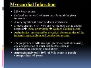
Myocardial Infarction. Death to myocardial tissue from prolonged ischemia >20”. Dead tissue is surrounded by area of ischemia. Etiology: Coronary thrombosis Coronary artery spasm Decreased O2 from blood loss, anemia, or low BP


Myocardial Infarction
E N D
Presentation Transcript
Myocardial Infarction • Death to myocardial tissue from prolonged ischemia >20”. Dead tissue is surrounded by area of ischemia. Etiology: • Coronary thrombosis • Coronary artery spasm • Decreased O2 from blood loss, anemia, or low BP • Increased O2 demand from rapid HR, thyrotoxicosis or cocaine abuse.
Classifications of MI • Degree of damage-full thickness or subendocardial (partially thru layers) • Location-anterior, inferior, posterior. Anterior wall of LV is most common from LADCA. • Point in time-acute, evolving, old
Manifestations CP more severe than angina Unrelieved by NTG & rest Dizziness, confusion, denial Diaphoresis, NV, rales, dyspnea, pallor Brady or tachycardia, gallop rhythm, decreased pulses Hypertension then hypotension Feeling of impending doom Diagnostic Tests ECG-elevation then depression of ST segment, inverted T wave, Q wave Elevated CK-MB, serum troponin WBC 12-15,000 Elevated glucose Elevated ESR Assessment of MI
Management of MI • Phase I-Acute phase-first 24 hours • First 6 hours crucial to salvage of the myocardium • Thrombolytics-streptokinase, tPA within 6 hours • Emergent PTCA • Pain control with NTG, morphine, and O2 (keep O2 sat >90) • ECG and telemetry
Management of MI cont’d • Phase 1-rest of hospital stay • Telemetry • Pain control for episodes of pain including O2 prn • Some of the following: nitrates, anticoags, antiplatelets, antilipids, Bblockers, ACE inhibitors, Ca++channel blockers, stool softeners • Heart healthy diet • Rest and progressive activity (Phase 1 of structured rehab) • Patient and family education on disease process, meds, activity, diet, stress reduction, follow-up, when to call MD.
Management of MI cont’d • Phase 2-after discharge and up to 6 months • Continue meds, diet, progressive activity. Exercise program may be structured outpatient activity or can be at home and self-directed and is usually started after 4 weeks. • Follow-up txs may be scheduled on outpatient or inpatient basis and may include stress test, PCTA with stenting, CABG, IABP, VAD, pacer, or AICD. • Dealing with complications
Management of MI cont’d • Phase 3-long term • Self directed with activity, meds, diet • Counseling-stress, financial, sex therapy, chaplain • OT or PT • Nutritionist • Social worker • Dealing with complications
Complications of MI • Can happen at any phase • Dysrhythmias are most common and develop from ischemia which destabilizes cell membranes, causes cell damage, and lyte imbalances. RCA supplies SA and AV nodes, LADCA supplies bundle branches. • Heart failure due to <contractility and leading to PE • Pericarditis from heart muscle inflammation
Phase I Assess and relieve pain Physical assessment Assess risk factors Assess dx test results Administer meds Promote rest and comfort and relief of anxiety Progressive activity Patient education Prevent complications Phase 2 & 3 Periodic assessments Continue meds, activity, diet Encourage reduction of risk factors Patient education Education and prevention of complications Nursing Management of MI
Congestive Heart Failure • Inability of heart to meet body’s demands. • Risk factors: • Previous MI-weakens muscle • HTN-increases afterload in great vessels • Hx of pericarditis-scar tissue causes constriction • Dysrhythmias-affects pump action • Anemia-increases HR • Thyroid disease-increases HR and BP • Lyte imbalances-affects regularity and contractility • COPD-increases afterload in PA • Diabetes-constricts small arteries • Valvular disorders-causes leakage
Right-sided Congestion in right chambers Increase in CVP Increase in size of RV Backflow to vena cava Congestion in jugular veins, liver, lower extremities Left-sided Congestion in left chambers Increase in size of LV Backflow to pulmonary veins Congestion in lungs Congestive Heart Failure-Types
Manifestations: SOB, DOE, PND, orthopnea, dry hacking cough or hemoptysis Crackles in LL, wt gain, edema, liver enlarges, oliguria, JVD Fatigue, anorexia <SBP, >DBP, <PP, tachy, S3 gallop rhythm Diagnostic Tests CXR-fluid and heart enlargement ECG-can reveal hx of heart problems Echo-enlargement, valvular function, condition of great vessels ABGs, O2 sat Liver enzymes, BUN, creatinine Assessment of CHF
Management of CHF • Primarily pharmacologic • Cardiac glycosides-digoxin • Cardiac stimulants-dobutamine • Diuretics-furosemide • ACE inhibitors • Vasodilators-dipyridamole • Ca++channel blockers • 6 small meals of NAS diet with >calories and protein • Fowler’s position, O2, rest, stress reduction, I&O, daily wts, possible fluid restriction
Nursing Management of CHF • To improve CO and gas x-change • Assessment of heart, lungs, tissue perfusion, edema, VS, telemetry, labs, I&O, daily wts • Administer and monitor med tx, diet, fluid restriction • Balance rest and activity; Fowler’s, O2 • Monitor for complications: acute PE, dysrhythmias, multisystem failure • Provide emotional support • Pt and family education: meds, rest and activity, diet, fluid status, S&S to report