Download
1 / 42
420 likes | 446 Views
Learn about bones and cartilages, their types, functions, growth, and structure. Discover how they support, protect, and aid in movement. Explore the classification and markings of bones.

E N D
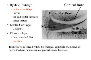
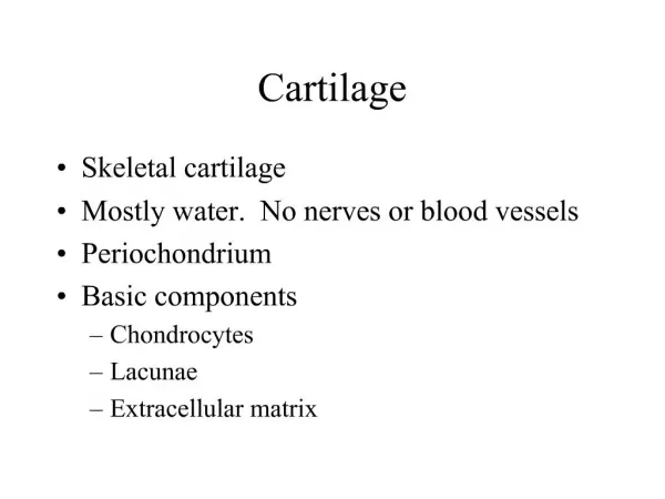
Skeletal Cartilage • Contains no blood vessels or nerves • Surrounded by the perichondrium (dense irregular connective tissue) that resists outward expansion • Three types – hyaline, elastic, and fibrocartilage
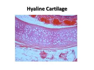
Hyaline Cartilage • Provides support, flexibility, and resilience • Is the most abundant skeletal cartilage • Is present in these cartilages: • Articular – covers the ends of long bones • Costal – connects the ribs to the sternum • Respiratory – makes up larynx, reinforces air passages • Nasal – supports the nose
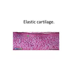
Elastic Cartilage • Similar to hyaline cartilage, but contains elastic fibers • Found in the external ear and the epiglottis
Fibrocartilage • Highly compressed with great tensile strength • Contains collagen fibers • Found in menisci of the knee and in intervertebral discs
Growth of Cartilage • Appositional – cells in the perichondrium secrete matrix against the external face of existing cartilage • Interstitial – lacunae-bound chondrocytes inside the cartilage divide and secrete new matrix, expanding the cartilage from within • Calcification of cartilage occurs • During normal bone growth • During old age
Bones and Cartilages of the Human Body Figure 6.1
Classification of Bones • Axial skeleton – bones of the skull, vertebral column, and rib cage • Appendicular skeleton – bones of the upper and lower limbs, shoulder, and hip
Classification of Bones: By Shape • Long bones – longer than they are wide (e.g., humerus) Figure 6.2a
Classification of Bones: By Shape • Short bones • Cube-shaped bones of the wrist and ankle • Bones that form within tendons (e.g., patella) Figure 6.2b
Classification of Bones: By Shape • Flat bones – thin, flattened, and a bit curved (e.g., sternum, and most skull bones) Figure 6.2c
Classification of Bones: By Shape • Irregular bones – bones with complicated shapes (e.g., vertebrae and hip bones) Figure 6.2d
Function of Bones • Support – form the framework that supports the body and cradles soft organs • Protection – provide a protective case for the brain, spinal cord, and vital organs • Movement – provide levers for muscles
Function of Bones • Mineral storage – reservoir for minerals, especially calcium and phosphorus • Blood cell formation – hematopoiesis occurs within the marrow cavities of bones
Bone Markings • Bulges, depressions, and holes that serve as: • Sites of attachment for muscles, ligaments, and tendons • Joint surfaces • Conduits for blood vessels and nerves
Bone Markings: Projections – Sites of Muscle and Ligament Attachment • Tuberosity – rounded projection • Crest – narrow, prominent ridge of bone • Trochanter – large, blunt, irregular surface • Line – narrow ridge of bone
Bone Markings: Projections – Sites of Muscle and Ligament Attachment • Tubercle – small rounded projection • Epicondyle – raised area above a condyle • Spine – sharp, slender projection • Process – any bony prominence
Bone Markings: Projections – Projections That Help to Form Joints • Head – bony expansion carried on a narrow neck • Facet – smooth, nearly flat articular surface • Condyle – rounded articular projection • Ramus – armlike bar of bone
Bone Markings: Depressions and Openings • Meatus – canal-like passageway • Sinus – cavity within a bone • Fossa – shallow, basin-like depression • Groove – furrow • Fissure – narrow, slit-like opening • Foramen – round or oval opening through a bone
Gross Anatomy of Bones: Bone Textures • Compact bone – dense outer layer • Spongy bone – honeycomb of trabeculae filled with yellow bone marrow
Bone Markings Table 6.1
Structure of Long Bone • Long bones consist of a diaphysis and an epiphysis • Diaphysis • Tubular shaft that forms the axis of long bones • Composed of compact bone that surrounds the medullary cavity • Yellow bone marrow (fat) is contained in the medullary cavity
Structure of Long Bone • Epiphyses • Expanded ends of long bones • Exterior is compact bone, and the interior is spongy bone • Joint surface is covered with articular (hyaline) cartilage • Epiphyseal line separates the diaphysis from the epiphyses
Structure of Long Bone Figure 6.3
Structure of Long Bone Figure 6.3a
Structure of Long Bone Figure 6.3b
Structure of Long Bone Figure 6.3c
Bone Membranes • Periosteum – double-layered protective membrane • Outer fibrous layer is dense regular connective tissue • Inner osteogenic layer is composed of osteoblasts and osteoclasts • Richly supplied with nerve fibers, blood, and lymphatic vessels, which enter the bone via nutrient foramina • Secured to underlying bone by Sharpey’s fibers
Bone Membranes • Endosteum – delicate membrane covering internal surfaces of bone
Structure of Short, Irregular, and Flat Bones • Thin plates of periosteum-covered compact bone on the outside with endosteum-covered spongy bone (diploë) on the inside • Have no diaphysis or epiphyses • Contain bone marrow between the trabeculae
Structure of a Flat Bone Figure 6.4
Location of Hematopoietic Tissue (Red Marrow) • In infants • Found in the medullary cavity and all areas of spongy bone • In adults • Found in the diploë (between inner and outer walls) of flat bones, and the head of the femur and humerus
Microscopic Structure of Bone: Compact Bone • Haversian system, or osteon – the structural unit of compact bone • Lamella – weight-bearing, column-like matrix tubes composed mainly of collagen • Haversian, or central canal – central channel containing blood vessels and nerves • Volkmann’s canals – channels lying at right angles to the central canal, connecting blood and nerve supply of the periosteum to that of the Haversian canal
Microscopic Structure of Bone: Compact Bone • Osteocytes – mature bone cells • Lacunae – small cavities in bone that contain osteocytes • Canaliculi – hairlike canals that connect lacunae to each other and the central canal
Microscopic Structure of Bone: Compact Bone Figure 6.6a, b
Microscopic Structure of Bone: Compact Bone Figure 6.6a
Microscopic Structure of Bone: Compact Bone Figure 6.6b
Microscopic Structure of Bone: Compact Bone Figure 6.6c
Chemical Composition of Bone: Organic • Osteoblasts – bone-forming cells • Osteocytes – mature bone cells • Osteoclasts – large cells that resorb or break down bone matrix • Osteoid – unmineralized bone matrix composed of proteoglycans, glycoproteins, and collagen
Chemical Composition of Bone: Inorganic • Hydroxyapatites, or mineral salts • Sixty-five percent of bone by mass • Mainly calcium phosphates • Responsible for bone hardness and its resistance to compression
Bone Development • Osteogenesis and ossification – the process of bone tissue formation, which leads to: • The formation of the bony skeleton in embryos • Bone growth until early adulthood • Bone thickness, remodeling, and repair
Formation of the Bony Skeleton • Begins at week 8 of embryo development • Intramembranous ossification – bone develops from a fibrous membrane • Endochondral ossification – bone forms by replacing hyaline cartilage
Intramembranous Ossification • Formation of most of the flat bones of the skull and the clavicles • Fibrous connective tissue membranes are formed by mesenchymal cells